TABLE OF CONTENTS
Vaginal Exfoliative Cytology (VEC)
Vaginal Exfoliative Cytology (VEC) in dogs is a diagnostic technique used to evaluate the reproductive status of female dogs and reproductive disorders.
Changes in exfoliate vaginal cells observed in simple and rapidly prepared smears can provide direct evidence of the progression of follicular development and estrogen secretion during proestrus, a reasonable estimate of the fertile period during oestrus and fairly accurate estimation of the end of the fertile period and the timing of subsequent events of a resulting pregnancy.
Vaginal Anatomy
The vaginal lining in anoestrus is only a few layers thick, relatively fragile and close to the blood supply. Increasing estrogen concentrations of proestrus cause rapid multiplication in the number of cell layers lining the vaginal vault.
The increased cell layers place the luminal lining cells further and further away from blood supply, resulting in the death of those cells. Oestrus is usually associated with more number of dead epithelial cells in the vaginal lumen.
Collection of Sample
Smears are obtained from the mid or cranial vagina using cotton swab technique. The lips of the vulva are gently separated with one hand and with the other hand cotton tipped applicator moistened with saline is introduced into the vagina in a cranio dorsal direction. The applicator is then rotated a complete revolution in each direction and withdrawn.
Preparation of Slide
The cotton tip is gently rolled from one end of a glass microscopic slide to the other. Do not rub or smear the cotton on the slide. Usually two slides should be prepared. The slides are air dried, dipped once or twice in methanol to prevent cellular deterioration or distortion.
Staining of Slide
Slides can be stained with Wright’s stain, Wright’s Giemsa Stain, Diff-Quick Stain, Freshly prepared Methylene blue, PAP or Haematoxylin-Eosin stain.
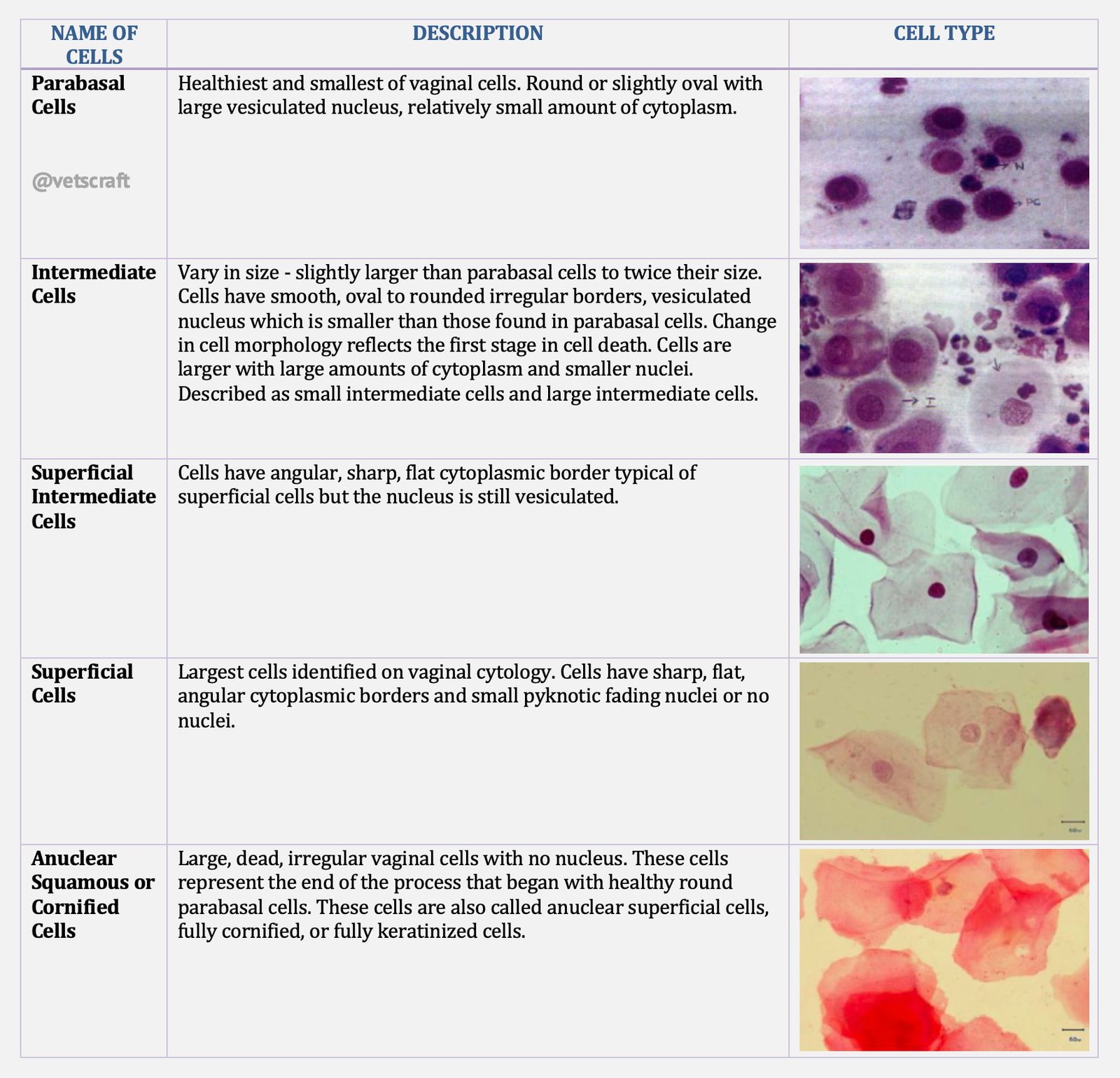
Examination of Slide
The different cell types represent stages of cell death. As healthy round vaginal cells die, they then become larger and more irregular in shape.
The nuclei within vaginal epithelial cells also undergo changes that reflect cell death. The nucleus becomes progressively smaller, then pyknotic, before disintegrating, leaving a nuclear “ghost” and then anuclear cell.
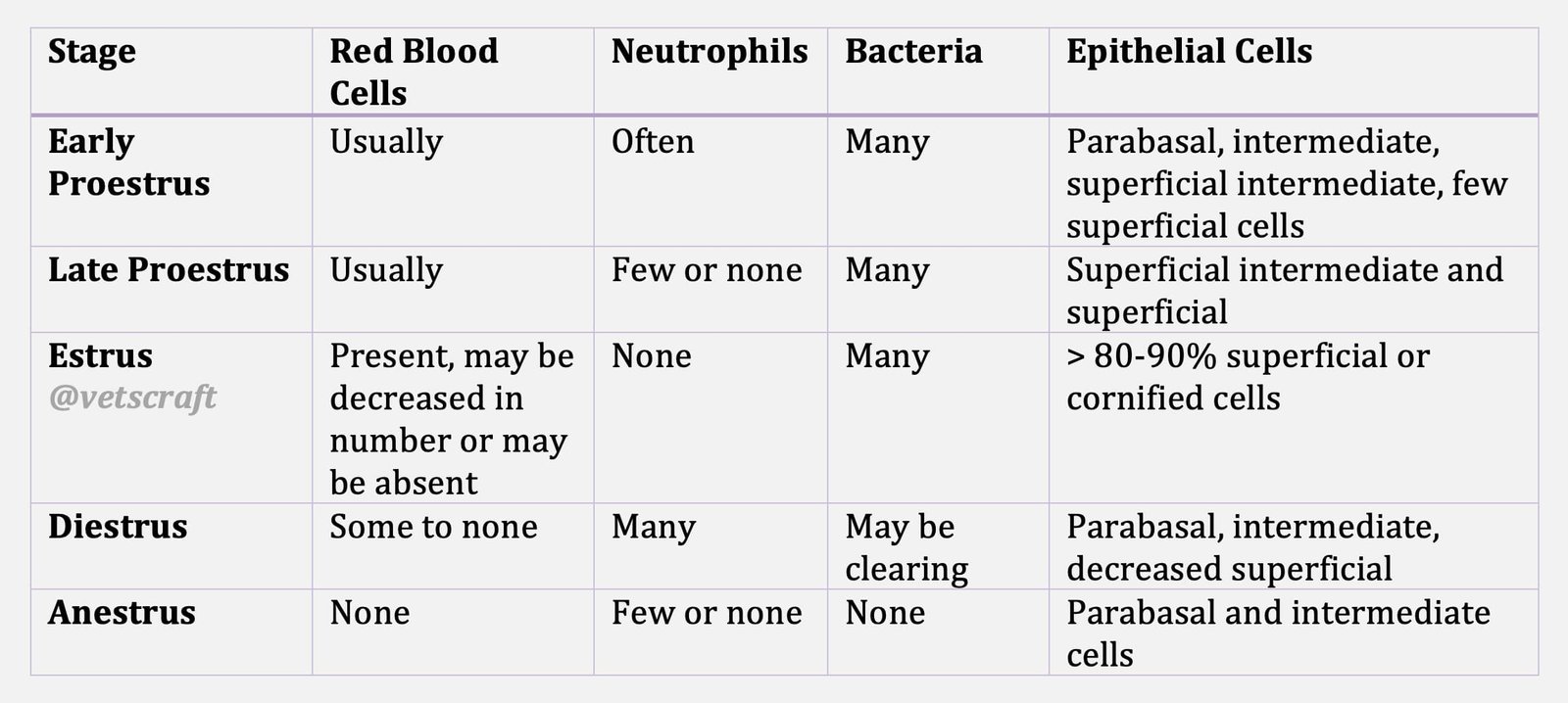
Cytological Stages of Estrous Cycle
Clinical Usefulness of Vaginal Cytology
Management of Normal Breeding
When the smear shows >80% of superficial or cornified epithelial cells, the female should be bred. Breeding should be done once every second, third or fourth day of estrus until the bitch refuses to breed.
Breeding Unusual Cycles
Some bitches fail to stand for a male because they are not exposed to the stud during standing heat (i.e. standing heat is not recognized because it does not occur between days 9 and 12 of the cycle). Vaginal cytology will help to identify the onset of standing oestrus.
Predicting Whelping Dates
Vaginal cytology is a superb tool in predicting the approximate whelping date. Identification of Day 1 of diestrum will help since whelping occurs near day 57 of diestrus.
Infertility Problems
Vaginal cytology is a crude reflection of plasma estrogen concentrations. Infertility cases should be evaluated with vaginal cytology and serum progesterone to find out if the problem is one of mismanagement, inadequate estrogen (poor follicular development) or some less common disorder.
Follicular Cysts
A follicular cyst is one which secretes estrogen. A bitch with follicular cyst will have prolonged proestrus or estrus. This could be confirmed with vaginal cytology followed by ultra sonography.
Vaginitis
An abnormally large number of healthy to degenerated neutrophils will be seen in smears of bitches with vaginitis.
Vaginal Tumors
Tumor cells of Transmissible venereal tumours and Transitional cell carcinomas of bladder could be identified.
Determining whether an abortifacient should be administered or not. The presence of spermatozoa or heads of spermatozoa together with superficial or cornified cells on vaginal cytology can be used to confirm whether breeding has taken place or not.

