TABLE OF CONTENTS
Vaginal Endoscopy in Dogs
Vaginal endoscopy in dogs is a minimally invasive diagnostic procedure used to examine the vaginal and reproductive tract.
This technique involves inserting a specialised endoscope into the vaginal canal, allowing for direct visualization of the mucosal lining, cervix, and other structures.
It is commonly used for diagnosing reproductive disorders, detecting infections, evaluating estrous cycle stages, and assisting in artificial insemination.
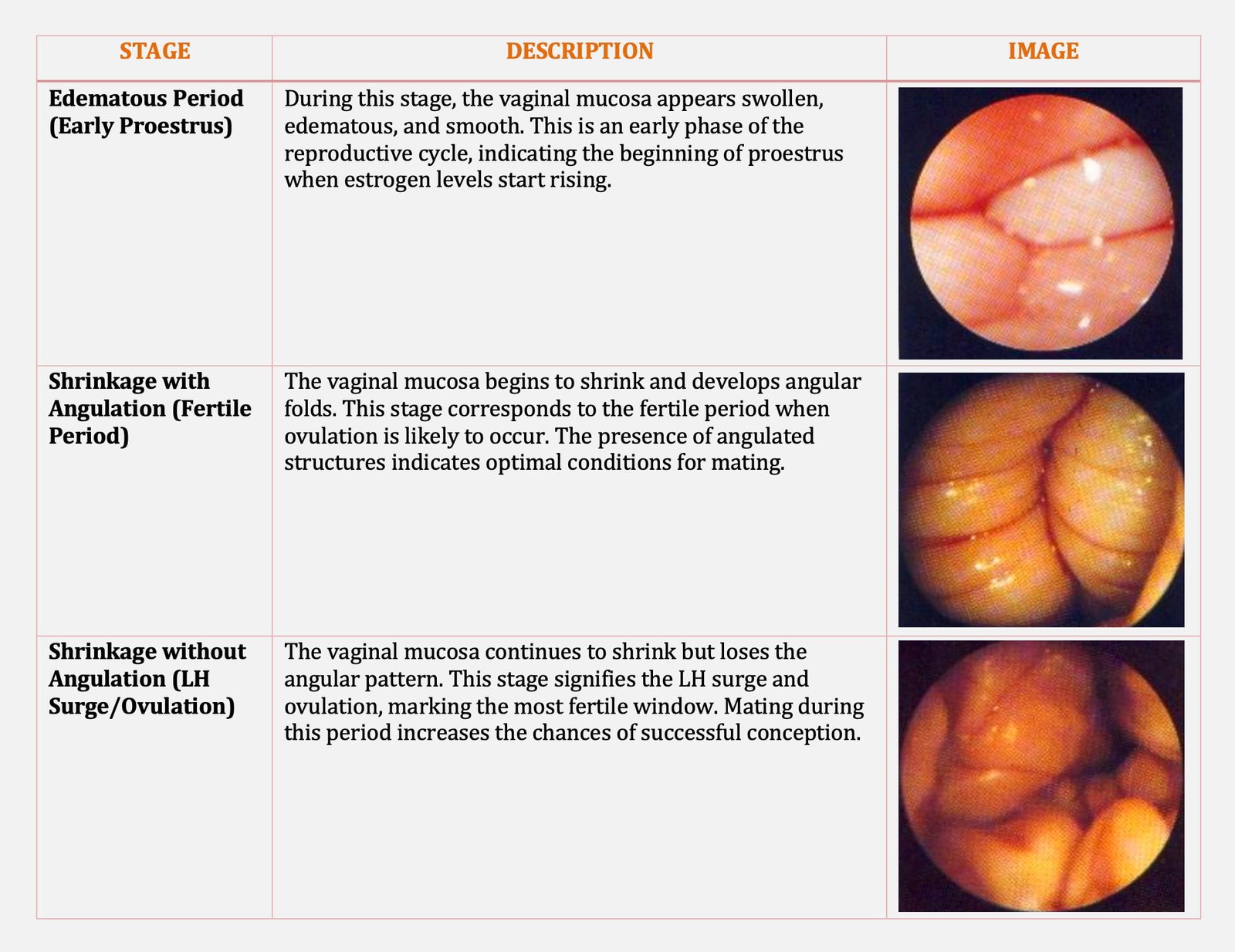
Vaginoscopy can be used to aid in timing natural breeding. Vaginal mucosa in proestrus appears rounded and oedematous. “Wrinkling” or “Crenulation” of mucosa is associated with LH surge. This is the time to begin breeding.
Breeding should be continued throughout the phase of maximal mucosal crenulation, seen as angulated folds of vaginal mucosa with sharp profiles. Breeding should be discontinued when the vaginal mucosa again becomes flaccid and smooth, with patchy red and white surface which is observed 6 to 10 days following LH surge.
Uses of Vaginal Endoscopy in Dogs
- Estrous cycle evaluation
- Artificial insemination assistance
- Diagnosis of reproductive disorders
- Detection of infections and inflammation
- Visualization of the cervix
- Foreign body detection and removal
- Tumor and polyp detection
- Biopsy collection
- Postpartum examination
- Assessment of surgical outcomes

