TABLE OF CONTENTS
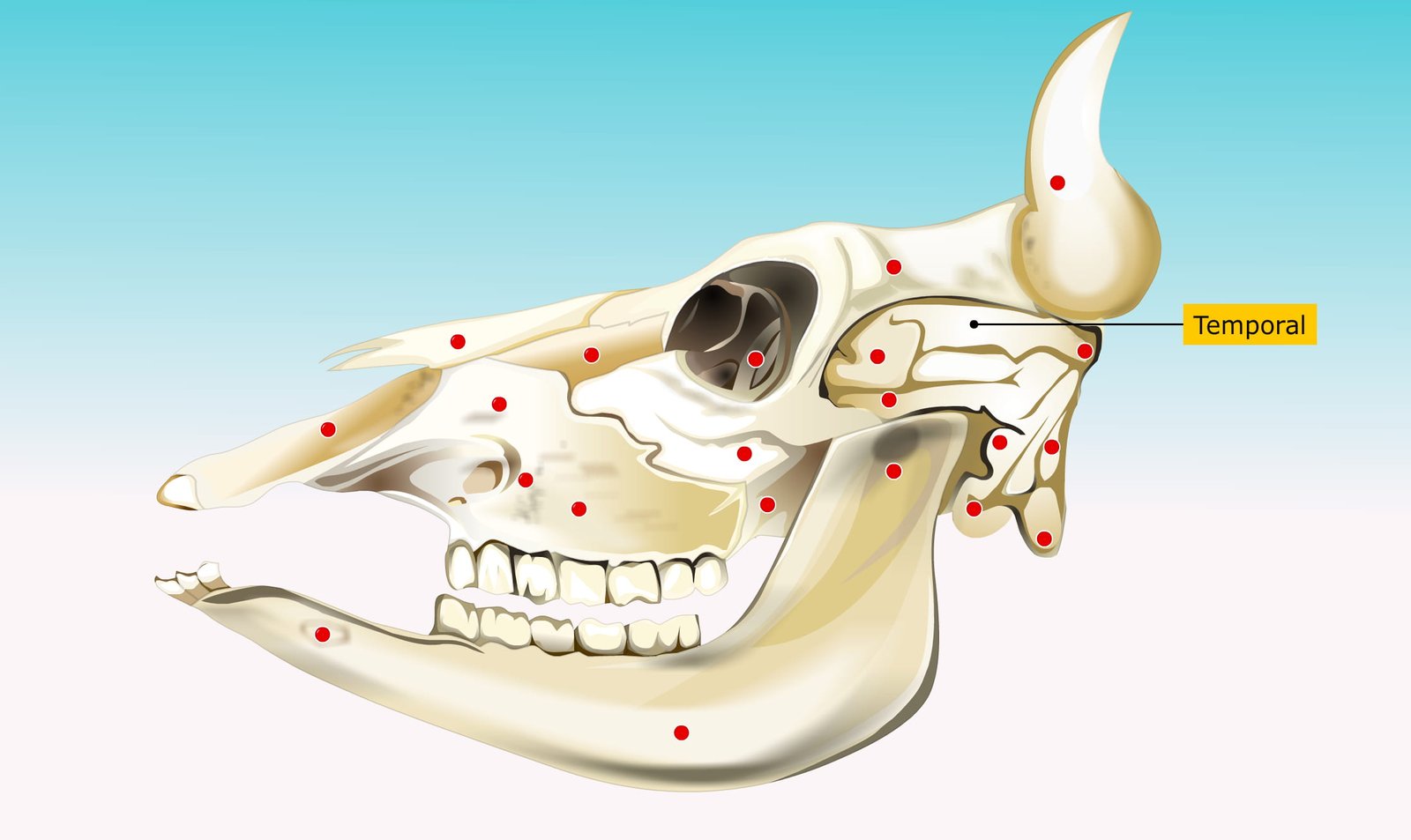
Temporal Bone in Domestic Animals (Ox, Horse, Dog, Fowl) – Anatomy & Comparative Structure
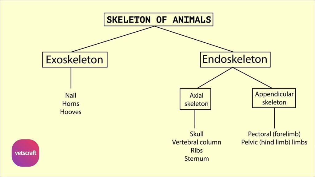
The temporal bone forms part of the lateral wall of the cranium and consists of squamous and petrous parts. It varies in form across domestic animals such as ox, horse, dog, and fowl.
Temporal Bone of Ox
The temporal bone forms part of the lateral walls of the cranium. It is situated between the occipital and parietal behind, and the frontal dorsally, and the sphenoid ventrally and medially.
Each temporal consists of squamous and petrous parts, which are completely fused at birth.
Squamous Temporal
The squamous temporal has a body and a zygomatic process. The external surface of the body is divided by the temporal crest into two parts.
The crest is continuous with the parietal crest above, turns forward below, and ends in a tubercle above the external acoustic meatus. This corresponds to the mastoid process (on the petrous part in the horse).
The area in front of the crest is the temporal fossa and presents two or three foramina leading into the temporal canal. The area behind the crest is smaller and meets the occipital bone.
The internal surface is overlapped by the parietal and sphenoid bones.
The zygomatic process is wide behind and narrow in front. The anterior part of the zygomatic process meets the zygomatic process of the malar bone to form the zygomatic arch. At its upper part, it forms dorsally part of the temporal fossa, and ventrally it presents an articular area—a condyle, a glenoid cavity, and a postglenoid process—for articulation with the mandible.
Behind the postglenoid process is the postglenoid foramen, which is the external opening of the temporal canal. This canal is formed by the opposition of the squamous and petrous parts of the temporal bone. It is a continuation of the groove mentioned in the squamous part of the occipital bone, and its internal opening lies above and behind the petrous part of the temporal bone. It contains the dorsal cerebral vein, which is a continuation of the transverse sinus of the dura mater.
Petrous Temporal
The petrous temporal is situated between the occipital bone behind and the squamous temporal in front. It consists of petrous and tympanic parts.
1. Petrous Part
- The petrous part contains the internal ear.
- The medial face is smooth and forms the lateral wall of the cerebellar compartment of the cranial cavity.
- It presents the internal acoustic (auditory) meatus for the VII and VIII cranial nerves.
- The fundus of the meatus is divided by a crest into two fossae. In the superior fossa is the origin of the facial canal (aquaeductus fallopius), which curves through the bone and opens externally at the stylomastoid foramen. It transmits the seventh cranial nerve.
- The inferior fossa presents small foramina for the passage of fibers of the eighth cranial nerve.
- Behind the meatus and near the posterior margin of the surface is the slit-like opening of the aquaeductus vestibuli, covered by a scale of bone. Below, it forms part of the cerebral compartment.
- The petrosal crest separates these two surfaces.
2. Tympanic Part
- The tympanic part is external and presents the following:
- The external auditory process is a curved plate of bone projecting through a notch in the squamous temporal and enclosing the external acoustic meatus.
- Below the external auditory process is a rod of bone, the hyoid process. This is connected by a bar of cartilage to the styloid cornu of the hyoid bone.
- Between the hyoid and the paramastoid processes is the stylomastoid foramen, which is the external opening of the facial canal.
- The bulla tympanica (auditory bulla) contains a cavity that forms part of the middle ear.
- The muscular (styloid) process of the petrous temporal arises from below the bulla for muscular attachment.
- Lateral to the root of this process is the petrotympanic (Glaserian) fissure for the chorda tympani nerve, and medially is a groove or semicanal called the osseous eustachian tube (auditory tube).
Comparative Anatomy of Temporal Bone
Comparative anatomy of the temporal bone refers to the study of structural similarities and differences in the temporal bone among various domestic animals, such as ox, horse, dog, and fowl.
Horse
- The squamous part is relatively larger. It presents a zygomatic process and a posterior triangular process in horse.
- The zygomatic process meets not only the malar but also the supraorbital process of the frontal.
- The posterior process arises from the posterior aspect of the body and presents a temporal crest on its lateral face. Its medial surface forms the outer boundary of the parietotemporal canal.
- The parietal bone contributes to the formation of the temporal canal (parietotemporal canal). There is a well-developed mastoid process between the squamous temporal and the paramastoid processes.
- The stylomastoid foramen lies between the hyoid and mastoid processes. The auditory bulla is smaller.
Dog
- The parts of the temporal bone fuse early in dogs.
- The zygomatic process is strongly curved.
- Its anterior part is beveled ventrally and articulates extensively with the corresponding process of the malar.
- The articular area is represented by a transverse groove, which extends onto the front of the large postglenoid process.
- A distinct mastoid process is present.
- The external acoustic meatus is very short and wide, so in the dry skull one can see into the tympanum.
- The bulla tympanica is very large, rounded, and smooth. Its medial side is united to the basilar part of the occipital bone.
- Above this junction, and roofed in by the union of the petrous parts and the basioccipital, is the petrobasilar canal. This canal transmits a vein from the floor of the cranium to the foramen lacerum posterius. The latter opens into a depression behind the tympanic bulla and transmits the 9th, 10th, and 11th cranial nerves.
- The carotid canal branches off from the petrobasilar canal, passes forward lateral to it through the medial part of the bulla tympanica, and opens in front of the carotid foramen. It transmits the internal carotid artery. The eustachian opening is immediately lateral to the carotid foramen. The muscular and hyoid processes are very rudimentary.
- The petrosal crest is sharp and projects prominently into the cranial cavity. The medial surface presents a deep floccular fossa above the internal acoustic meatus. The anterior angle is perforated by a foramen for the trigeminal nerve.
Fowl
- The squamous temporal lies on the lateral aspect of the cranium. It presents the supraorbital and the zygomatic processes in fowl.
- The former is fused to that of the frontal and furnishes a facet for the quadrate bone.
- The petrous temporal is concealed by the adjacent bones. It forms the floor of the tympanic cavity and part of the facet for the quadrate bone.