TABLE OF CONTENTS
Anatomy of Tarsal Bones in Animals: Ox, Horse, Sheep, Goat, Pig, Dog, Rabbit, and Fowl
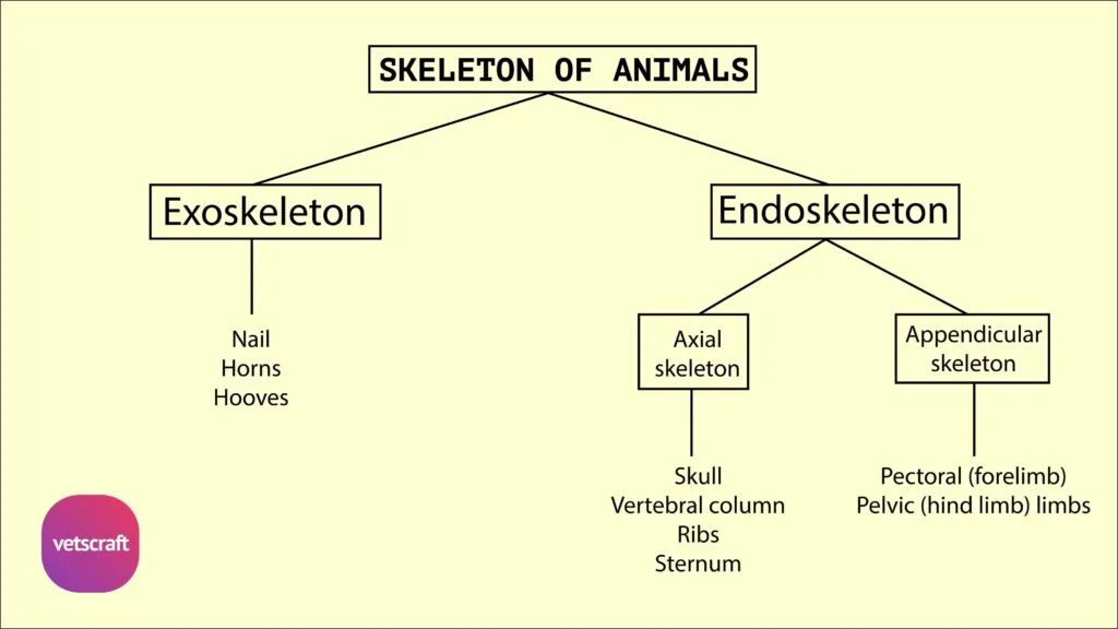
Tarsal bones are a group of small, irregular bones that form the hock joint or ankle region in animals. These bones connect the lower leg (tibia and fibula) to the metatarsal bones of the foot and play a crucial role in supporting body weight, providing flexibility, and enabling movement of the hindlimb.
The number, fusion pattern, and arrangement of tarsal bones vary across species like ox, horse, dog, pig, and fowl, making them an important feature in comparative anatomy.

Tarsal Bones of Ox
The tarsal bone is composed of five short bones in the ox.
- Tibial Tarsal (Astragalus)
- Fibular Tarsal (Os Calcis)
- Fused Central and Fourth Tarsal (Scapho-Cuboid)
- First Tarsal (Cuneiform Parvum)
- Fused 2nd and 3rd Tarsal (Cuneiform Magnum)


1. Tibial Tarsal (Astragalus)
- The tibial tarsal (astragalus) has six surfaces.
- The proximal and dorsal faces are continuous and form a trochlea with vertical ridges for the tibia and the lateral malleolus.
- The distal face articulates with the fused central and fourth tarsals.
- The plantar face corresponds to the fibular tarsal. A narrow, elongated area on the medial aspect of the lower part of this face is for the plantar prolongation of the fused central and fourth tarsals.
- The lateral face is more depressed and shows two facets for the fibular tarsal.
2. Fibular Tarsal (Os Calcis)
- Fibular tarsal (os calcis) is the largest tarsal bone, is placed behind and lateral to the tibial tarsal.
- It has a body and a medial process—the sustentaculum tali.
- The lateral face of the body is flat and rough, while the medial face presents two facets for the tibial tarsal.
- The process, the sustentaculum tali, projects medially and presents a facet dorsally for the tibial tarsal.
- The body is prolonged above to form the tuber calcis.
3. Fused Central and Fourth Tarsal (Scapho-Cuboid)
- The proximal face presents two concave areas for the tibial tarsal, a projection postero-medially for the narrow medial facet on the distal face of the tibial tarsal, and a narrow facet on its lateral aspect for the fibular tarsal.
- The distal face is uneven. The medial half is higher in level and presents a large facet in front for the second and third tarsals, and a small facet behind for the first tarsal. The lateral half presents two facets separated by a transverse groove and articulates with the large metatarsal bone.
4. First Tarsal (Cuneiform Parvum)
- The first tarsal (cuneiform parvum) is a small, round piece of bone situated at the postero-medial aspect of the tarsus.
- The proximal face articulates with the fused central and fourth tarsal bones.
- The distal face articulates with the large metatarsal, and the dorsal face with the fused second and third tarsals.
5. Fused 2nd and 3rd Tarsal (Cuneiform Magnum)
- The fused 2nd and 3rd tarsal (cuneiform magnum) is placed beneath the fused central and 4th tarsal on its medial aspect.
- The proximal face is concavo-convex from front to back and articulates with the fused central and 4th tarsal.
- The distal face articulates with the large metatarsal.
- The plantar face has a facet for the first tarsal.
- The dorsal and medial faces are continuous, convex, and rough.
Comparative Anatomy of Tarsal Bones
The tarsal bones show significant variation among domestic animals in both number and arrangement.
Sheep and Goat
The tarsal bones of sheep and goats are similar to those of the ox.
Horse
The horse has six tarsal bones.

- In the tibial tarsal, the ridges of the trochlea curve obliquely downward and outward. The lateral face has no facets for the fibular tarsal.
- The fibular tarsal is short and thick. It does not articulate with the lateral malleolus. The medial process is larger.
- The central tarsal (scaphoid) is not fused to the fourth tarsal (cuboid). It is flat and irregularly quadrilateral. The proximal face is concave and articulates with the tibial tarsal. The distal face is convex and articulates with the third tarsal and the fused 1st and 2nd tarsal. The dorsal and medial borders are continuous and rough. The lateral border is oblique and has two facets for the cuboid.
- The fused 1st and 2nd tarsal (cuneiform parvum) is irregular in shape. The medial face is convex. The lateral face is concave. The proximal face is concave and has two facets for the central tarsal. The distal face articulates with the large and the medial small metatarsals. The plantar end is prolonged downward into a nodular projection.
- The third tarsal (cuneiform magnum) is triangular in outline. The proximal face is concave and articulates with the central tarsal. The distal face is convex and is for the large metatarsal. The medial border has no facet for the fused 1st and 2nd tarsal, and the lateral border has two facets for the fourth tarsal.
- The fourth tarsal (cuboid) has six surfaces. It is not fused to the central tarsal. The proximal face is transversely convex and is for the fibular tarsal and the tibial tarsal. The distal face presents two facets separated by a sagittal ridge for the lateral small metatarsal bones. The medial face has facets for the central and third tarsals. The dorsal, lateral, and plantar surfaces are convex and rough.
Pig
The pig has seven tarsal bones.

Dog
The dog has seven tarsal bones.

Rabbit
The rabbit has six tarsal bones.

Fowl
- The tarsal bone is absent as such in the adult fowl.
- In the proximal row, the embryonic elements fuse with the tibia, and in the distal row, with the metatarsus.