TABLE OF CONTENTS
Surgical affections of udder and teats
Surgical affections of udder and teats are Supernumerary or extra teats, Bovine ulcerative mammitis (sore teats), Udder and teat abscess, Teat laceration and fistula, Lactolith (milk stone), Teat canal polyp, Teat spider, Fibrosis of teat canal, Tumour of mammary gland, Teat stenosis (Hard milker), Teat leaker (Free milker), Blind teats etc.
Surgical affections of udder and teats in animals are-
- Supernumerary or extra teats
- Bovine ulcerative mammitis (sore teats)
- Udder and teat abscess
- Teat laceration and fistula
- Lactolith (milk stone)
- Teat canal polyp
- Teat spider
- Fibrosis of teat canal
- Tumor of mammary gland
- Teat stenosis (Hard milker)
- Teat leaker (Free milker)
- Blind teats
Surgical affections of udder and teats are getting much attention now a days as these affects the economy of the farmer. Milk alone contributes around 63% to the total output from livestock.
The udder and teats are vulnerable to external trauma or injury because of their anatomical location, increase in size of udder and teats during lactation, faulty methods of milking, repeated trauma to the teat mucosa, injury by teeth of calf, unintentionally stepped on teat, paralysis resulting from metabolic disturbances at parturition.
Any disease condition of udder and teats not only causes painful milking but also makes udder and teats prone to mastitis. The diseases of udder can be congenital anomalies are known at the time of first calving but acquired anomalies can affect any stage of lactation.
Supernumerary or extra teats
Supernumerary or extra teats are often seen on the posterior surface of udder and in-between the teat. They may be functional or nonfunctional, functional activity can be determined only after parturition of the animal.
They frequently interfere with free milking process and are objectionable on show animals.
It has been reported that presence of supernumerary teats has no significant effect on milk yield, lactation length, age at calving, conception rate and service period.
Surgical removals of these teats are best in young animals and in case of older cow in dry condition. Surgery performed under local infiltration analgesia with two elliptical incisions at the junctions of teat and udder and skin wound closed with interrupted suture using nonabsorbable suture material.
Bovine ulcerative mammitis (sore teats)
The teats become painful due to presence of crakes, traumatic injuries, lesions due to disease conditions such as pox, FMD etc. If these lesions are not treated well in time, the animal will not allow touching the affected teat for milking.
These lesions become ulcers in due cource of time and the condition are then known as bovine ulcerative mammitis.
Oozing of blood from injured teat causes contamination of milk while milking thereby making it unfit for human consumption.
In such cases, sterilized teat siphon should be used to drain the milk out. For treatment of such painful lesions, the wound should be washed with light potassium permanganate solution and then soothing preparation such as iodized glycerin, bismuth iodoform paraffin paste, zinc oxide ointment or antiseptic dressing with soothing emollient may be continued till the complete healing of the lesion occurs.
Udder and teat abscess
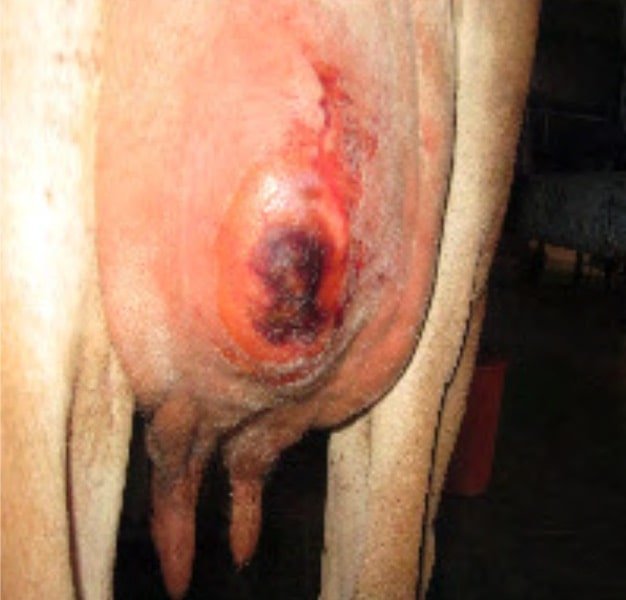
Abscess formation occurs more often on the udder than the teat. Many cases with chronic mastitis especially due to resistant microbes suddenly develop abscessation on side of affected udder. Such cases can easily be diagnosed by puncturing the swollen part.
The abscess cavity is opened for complete drainage of pus. After drainage of the pus, the cavity is dressed with tincture iodine followed by application of soothing agents until obliteration of abscess cavity.

In case of necrosis of teat or udder, amputation of teat or affected quarter is recommended followed by daily dressing till complete healing of wound occurs.
Teat laceration and fistula
The Teat laceration and fistula is mostly observed in those animals that have long teats and pendulous udder.
When animal tries to jump over the barbed wire or pass through the thorny bushes, their teat get teared due to laceration of skin and muscles. If this laceration is deeper, then even teat canal gets opened and milk will start flowing through the teared portion. This condition is called as teat fistula.
The cases of teat fistula are considered as emergency because any delay in repair of such teat will cause development of mastitis or necrosis of the teat. For repair of such teat, all aseptic precautions should be taken into considerations.
A full coverage of systematic antibiotic is required and for proper drainage Larson’s teat plug is used. Different suture techniques are used to repair the teat fistula but double layer simple continuous suturing with PGA 3/0 and in between simple vertical mattress simple interrupted suturing of skin with nylon 1/0 is found suitable for repair of teat fistula.
Surgical technique for teat fistula
After local infiltration or ring block by local anaesthetics-
Moussu’s method
The edges of the teat fistula are freshened and are sutured by a set of mattress sutures passing through the skin and subcutis on one edge and only subcutis on the other edge.
Another layer of interrupted sutures are applied and a teat siphon is introduced and bandaged.
Gold’s method
Following freshening of the fistula a series of mattress sutures are placed through the muscular and skin of either side with out piercing the mucous edge.
Lactolith (milk stone)
Milk stone are formed into the teat canal when the milk is rich in minerals and salty in taste due to super saturation of salts.
The stone moves freely in teat canal and hinder the milk flow, if large in size.
They usually get washed out along with ilk but if large in size then it can be crushed with small forceps or cutting the sphincter with itchy teat knife or test bistoury and milked out.
Teat canal polyp
Teat canal polyp are small pea sized growths attached to the wall of teat canal. The polyps hinder the milking process and sometimes even block the passage of teat canal.
Teat polyps can easily take out by Huges teat tumour extractor. If its location is above the teat canal thelotomy is the best method for resection of excessive tissue.
Postoperative gentamicin and prednisolone infusion for five consecutive days found suitable to check infection as well as helpful in checking further growth of the polyp.
Teat spider
Teat spider condition is usually due to congenital absence of teat cistern or canal.
It can be acquired in cases of injury, tumour or inflammation of mammary tissue resulting in formation of thin or thick membrane, situated either at the base or middle of the teat.
This membranous obstruction removed by teat scissor, Huges teat tumour extractor, teat bistouries or Hudson spiral teat instrument.
Fibrosis of teat canal
Fibrosis of teat canal condition is commonly observed in most of the lactating animals where a hard fibrous cord like structure is observed in the teat.
Exact cause of this condition is not clear. However, repeated trauma due to mechanical injuries, thumb milking and calf suckling are the main contributory factors.
Sometimes mastitis can also result into fibrosis of quarter followed by teat canal. This fibrotic cord will obstruct the teat canal and will create hindrance during milking.
In such cases, initially hot water fomentation followed by counter irritant massage such as iodine ointment and turpentine liniment massage is very useful.
In some cases it is advisable to place polythene catheter after removal of fibroid mass by Hugs teat tumour extractor.
Tumor of mammary gland
Tumors of mammary gland are infrequently in lactating animals however, fibro adenoma reported in heifer.
The growth can be surgically removed under caudal block or local infiltration analgesia.
Teat stenosis (Hard milker)
Teat stenosis (Hard milker) is the condition when teat sphincter gets contracted due to repeated trauma resulting in hard milking of teat. During milking one has to apply more force to take the milk out and milk will come out in fine stream.
Stenosis of streak canal without acute inflammation can be treated successfully by incising the sphincter in three directions with teat knife, Bard parker blade No.11, Udall’s teat knife, McLean teat knife.
Teat leaker (Free milker)
Teat leaker (Free milker) condition is just reverse of teat stenosis. It can be due to injury or relaxation of teat sphincter.
In this case milk will go on leaking and sometimes infection may gain entry leading to mastitis. This condition is treated by injection of 0.25 ml of lugo’s iodine around the orifice or scarification and suturing with one or two stitches with monofilament nylon.
Blind teats
Blind teats condition may be congenital or acquired due to any trauma near the teat sphincter. Such cases generally reported just after parturition on palpation milk thrill found in teat cistern on pressing milk passed backward toward milk udder cistern.
Imperforated teat treated by 15 gauze needle, after creating opening, it is further dilated using hugs teat tumour extractor, milk canula fixed for 24 hour after that frequent milking advised at 4 to 6 hours intervals to prevent adhesion.
Administration of proper antibiotics is done for a minimum period of 3-5 days.