TABLE OF CONTENTS
Surgical affections of trachea and larynx
Surgical affections of trachea and larynx in animals are Tracheal Collapse, Tracheal Stenosis, Foreign body in trachea, Tracheal Rupture, Laryngeal Collapse, Laryngeal Paralysis, Everted laryngeal saccules, Laryngeal trauma, Laryngeal stenosis, Proliferative diseases of larynx etc.
Surgical affections of trachea and larynx are-
- Tracheal Collapse
- Tracheal Stenosis
- Foreign body in trachea
- Tracheal Rupture
- Laryngeal Collapse
- Laryngeal Paralysis
- Everted laryngeal saccules
- Laryngeal trauma
- Laryngeal stenosis
- Proliferative diseases of larynx
Tracheal Collapse
Tracheal Collapse condition is reported in all age group of dogs with an average of 7 years. Early signs are mild productive cough progressing to severe exercise intolerance.
Dyspnoea and harsh rales may be noticed. Abdominal lift is more prominent when thoracic tracheal collapse is severe.
Palpation, radiographs and fluoroscopy of cervical and thoracic region of trachea can be of diagnostic aid for Tracheal Collapse.
Surgical correction should not be attempted unless the upper respiratory obstruction, stenotic nares, laryngeal collapse are relieved.
Dorsal tracheal membrane plication, internal stents, tracheal ring transection and external support are the four common techniques used for correction of tracheal collapse.
Tracheal Stenosis
Tracheal stenosis is the abnormal narrowing of the trachea that reduce diameter of tracheal lumen.
Narrowing of the tracheal lumen can be due to formation of scar tissue because of endotracheal tube pressure, blunt or penetrating trauma, tracheostomy and tracheal anastomosis.
Dyspnea is observed during inspiration and expiration. Tracheoscopy can be used to visualise the changes in the lumen shape and mucosal surface.
Dilatation of stenosis is achieved by passing large rigid bronchoscope, stretching and flattening the stenosis.
Resection of long tracheal segments may require special techniques to allow anastomosis.
Resection of mucosal and submucosal granulation tissue and mature scar leaves a mucosal defect that leads to recurrence.
Foreign body in trachea
Small light objects may be inhaled deep within the trachea, which leads to chronic pneumonia, abscess and fistulous tracts.
Acute onset of cough and dyspnea is common in case of foreign body in trachea. and high frequency rales may be heard in partial obstruction.
Plain radiograph, bronchoscopy are the diagnostic aid to confirm foreign bodies in trachea.
The retrievable foreign body can be removed from with the help of rigid hollow bronchoscope or flexible fiber-optic endoscope.
Tracheal Rupture

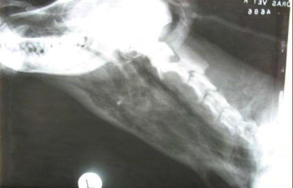
Tracheal Rupture in a dog (Right Lateral x-ray radiograph of neck)
Etiology
In small animals dog bitten wounds and penetrating trauma on the neck region might result in punctured wounds on the trachea.
Symptoms
- Emphysema
- Hissing sounds of air in the trachea

Subcutaneous emphysema following tracheal rupture in a dog
Treatment
Suturing of the punctured trachea or larynx with a monofilament absorbable suture material (1/0 PDS).
Laryngeal Collapse
Laryngeal Collapse occurs due to result of cartilage fracture or loss of supporting function of the cartilage. This is a brachycephalic airway syndrome and is a progressive disease.
Predisposing factors are stenotic nares and elongated soft palate. A temporary tracheostomy is necessary to ensure adequate passage of air during surgery and also for post operative recovery. Dogs with stenotic nares and elongated soft palate and everted laryngeal saccules are treated first.
Permanent tracheostomy is an alternative for dogs with severe laryngeal collapse even after resection of above mentioned conditions.
Laryngeal Paralysis
In dogs and cats, Laryngeal Paralysis usually occurs from an interruption of the innervation to the intrinsic muscles of the larynx.
Any disruption of normal nerve transmission of vagus or recurrent laryngeal nerves; may be either congenital or acquired.
Damage or severance of the laryngeal nerves subsequent to cervical surgery or trauma also cause paralysis.
Clinical Signs include change in voice followed by gagging and coughing in early stages. In severe cases, severe dyspnea, cyanosis or syncope can be noticed.
Unilateral or bilateral arytenoid cartilage lateralisation, ventricular cordectomy, and permanent tracheostomy are the surgical procedures used to correct laryngeal paralysis.
Everted laryngeal saccules
Everted laryngeal saccules mostly seen in brachycephalic breeds. The saccules evert in response to decrease in pressure that is created within the larynx during inspiration.
Everted tissue rapidly becomes edematous and partially occludes the ventral rima glottis.
The saccule is grasped with long Allis tissue forceps, the saccule is amputated at its base while applying rostral traction.
Laryngeal trauma
The intrinsic laryngeal trauma is caused by rough intubation for anesthesia and examination. Long term intubation can result in temporary laryngeal paralysis and aspiration.
Extrinsic trauma due to accident is uncommon. Submucosal hemorrhage, mucosal laceration, luminal obstruction due to cartilage mal-alignment or hematoma are signs of laryngeal damage.
Laryngoscopy and esophagoscopy are important methods of examining the injuries.
Advancement flaps of mucosa from the piriform area are used to cover rostral laryngeal cartilage surfaces covered with mucosa. Fractured cartilages are debrided, trimmed and closed with preplaced interrupted sutures.
Laryngeal stenosis
Obstruction of the larynx by granulation tissue and cartilage degeneration and collapse results in progressive reduction in airway diameter.
These lesions vary from web stenoses to broad based scar tissue covered by mucosa. Laryngeal stenosis is a complication of laryngeal surgery and trauma.
Proliferative diseases of larynx
Granulomatous laryngitis is a chronic inflammatory disease and the lesions are found around the arytenoid processes and cause stenosis. Regression of the lesion usually occurs with debulking of the mass and steroid theraphy.
Primary neoplasia of the larynx is rare in dogs and cats. Only Squamous cell carcinoma is the most common laryngeal neoplasia in small animals. Inflammatory polyps and laryngeal cysts are occasionally encountered in the laryngeal region.