TABLE OF CONTENTS
Surgical affections of the Eyes
Surgical affections of the Eyes are Tarsal cyst, Hordeolum, Dacryo-adenitis, Blepharitis, Entropion and Entropion, Lagophthalmos, Blepharospasm, Ptosis, Trichiasis and Distichiasis, Conjunctivitis, Prolapsed nictitans gland, Limbus melanoma, Naso lacrimal duct occlusion etc.
Anatomy of eye
The eyeball and its surroundings–
- The eyeball (Oculus bulbi) is situated within the bony cavity known as the orbit and is protected by the eyelids anteriorly. It is surrounded by muscles and a thick padding of retroubular fat posteriorly.
- The bony orbital rim is complete in some species. The term closed orbits is used when the bony orbital rim surrounding the eyeball is complete. Closed orbit is seen in man, horse, cattle and camel. An open orbit is an orbit with the bony rim incomplete so that part of it is made up of a fibrous ligament. Open orbit is seen in cat, elephant, pig, dog and birds.
- The anterior segment of the eye is the portion of the eye between the cornea and the lens consisting of the eyelids, conjunctiva, cornea, iris and pupil, and the anterior capsule of the lens. Posterior segment of the eye is from the lens backwards, namely vitreous and retina.
Visual function tests for eyes
A blind animal is nervous and is easily excitable. It shows anxious movements of the ears in an attempt to grasp the environment.
It walks with the head held upwards and takes very cautions steps and has a “feeling gait”.
During progression it stumbles on account of the inability to see obstacles on an uneven ground; and in order to avoid such obstacles it may lift the limbs unusually high (“high stepping”).
When driven towards an object like a wall or a post, the animal may go and strike against the object because of the inability to see.
When light is suddenly flashed into a normal eye, immediate closure of the eyelids is noticed. This is a protective reflex known as palpebral reflex. Palpebral reflex is absent in a blind eye.
Photomotor pupillary reflex (Photomotor pupillary reaction)
Photomotor pupillary reflex (Photomotor pupillary reaction) is the ability of the pupil to react to changes in light. If the eyes are normal, the pupil contracts when exposed to bright light and dilates when there is shade or darkness.
Absence of this reflex may indicate some abnormality.
Consensual reflex: If both eyes are visual, the flashing of light into one eye constricts both the pupils. This is called crossed reflex or consensual reflex. If one eye is blind, flashing of light into the blind eye will not induce pupillary reflex of the normal eye.
Detailed ophthalmic examination
Naked eye examination
Gross abnormalities of the anterior segment of the eye can be detected by naked eye examination with the aid of artificial illumination if necessary.
Using magnifying binocular loupe
The binocular loupe consists of two magnifying lenses and its use is therefore preferable to naked eye examination.
By using ophthalmoscope
Ophthalmoscope is mainly used to view the fundus.
It is necessary to dilate the pupil. This can be brought about by instilling a solution of homatropine (2%) or tropicamide into eye about fifteen to thirty minutes before ophthalmoscopic examination.
The ophthalmoscope contains lenses of varying powers through which the examination can be conducted. The anterior segment of the eye can be examined by using a lens ranging from +12 to +20. For observing the lens +8 to +12, and for vitreous humour 0 to +8, are required. For the fundus of the eye (retina, optic disc) – 3 or less, may be suitable.
By using tonometer (tonometry): The intraocular pressure (IOP) can be measured by using an instrument called tonometer.
There are two methods of tonometry, indentation tonometry using schiotz tonometer and applanation tonometry using Tonopen – Vet.

The normal intraocular pressure in the dog ranges from 16 to 30 mm of mercury. The normal IOP in man is 15 to 20 mm of mercury.
Schirmer tear test
The Schirmer tear test can be performed with commercially available Schirmer tear strips.
These strips have a notch at one end which is placed into the ventral conjunctival cul de sac.
The strip is allowed to remain in the cul de sac for exactly one minute.
The strip is removed after a minute and the distance the wetness have traveled down the strip is immediately measured in millimeters from a scale printed directly on the strip.
Normal values in the dog are 15 to 25 mm/minute.

Naso lacrimal flush
Naso lacrimal flush is the irrigation of the nasolacrimal duct system. Fluorescein dye is instilled on the eye.
Surgical affections of the Eyes
Major surgical affections of the eyes in animals are–
- Chalazion or Tarsal cyst
- Hordeolum or stye
- Dacryo-adenitis
- Blepharitis
- Entropion
- Ectropion
- Trichiasis and Distichiasis
- Ptosis (Blepharo ptosis)
- Lagophthalmos
- Blepharospasm
- Conjunctivitis
- Epiphora
- Symblepharon
- Ankyloblepharon
- Pterygium
- Neoplasms of the third eyelid
- Prolapsed nictitans gland
- Limbus melanoma
- Naso lacrimal duct occlusion
Chalazion or Tarsal cyst
Chalazion or Tarsal cyst is a cyst caused by the distension of a tarsal gland with secretion when it is inflammed. The size of the cyst may be about the size of a pea or more.
Treatment is incise and remove the contents of the cyst using a Chalazion forceps.
Hordeolum or stye
Hordeolum or stye is a localized inflammation of the hair follicles of the eye lashes due to staphylococcal infection.
Treatment
One or two neighboring eyelashes are plucked with foreceps so as to open the abscess and drain the pus. After draining the pus topical ophthalmic antibiotic eye ointment / drops are indicated.
Dacryo-adenitis
Dacryo-adenitis is the inflammation of lacrimal gland. treatment is Fomentations, antibiotics, etc. Do not open before it is mature. Spontaneous rupture and healing usually happens.
Blepharitis
Blepharitis or inflammation of eyelids, causes ulceration of the palpebral borders. The ulcers contain a yellowish or greyish sticky discharge. The eyelids may stick together.
Treatment for blepharitis is symptomatic and antibiotics are used to control infection.
Entropion
Entropion is may be congenital or acquired in animals. It is Inward deviation of the palpebral border, trichiasis, Distichiasis, etc.
Surgical correction is the only treatment. A fold of skin parallel to the affected palpebral border is held by a forceps, enough to cause the correction of the abnormality, and is severed and removed.
The wound is sutured by ordinary apposition sutures.
Ectropion
Ectropion is the outward deviation of the palpebral border resulting in an abnormal exposure of the conjunctiva.
Surgical correction is the only treatment. Site is 1⁄2 to 1 cm away from the free border of the eyelid for surgery.
A ‘V ‘ – shaped cutaneous incision is put with the base of the “V” close to the affected border of the lid.
The triangular flap of skin outlined is worked loose from its apex by undercutting to effect correction of palpebral border.
The gap thus caused at the apex is closed by suturing the sides of the “V” incision to form a “Y”.
Trichiasis and Distichiasis
In trichiasis the eyelashes are directed slightly inwards so that they irritate the cornea and conjunctiva. Distichiasis is a congenital condition in which two rows of eyelashes are noticed on each lid and the inner row causes irritation of the conjunctiva. Distichiasis supposed to be hereditary.
Treatment
- Epilation or plucking of the eyelashes
- Destroying the hair roots by eletrocautery
- Complete removal of the hair roots by snipping the inner border of the lid
- Operation for entropion may prevent the eyelashes irriating the cornea
Ptosis (Blepharo ptosis)
Ptosis (Blepharo ptosis) is the dropping of the upper eyelid may be congenital. It may be due to paralysis of the seventh cranial nerve.
The condition may be temporary and may become normal without treatment. Surgical correction when necessary can be done as for entropion.
Lagophthalmos
Lagophthalmos is s condition in which the eye cannot be completely closed (Lagos means hare).
Etiology
- Paralysis of the orbicularis oculi muscle resulting from injury to the seventh cranial nerve
- Prolapse of harderian gland
- Inflammed lacrimal gland
- Growth on the cornea.
- Staphyloma.
- Granulations on the edges of the eyelids.
- Lagophthalmos causes drying of the cornea and conjunctiva
Treatment
Remove the cause. Ophthalmic lubricants in the form of gel may be instilled at frequent intervals to moisten the cornea and conjunctiva.
The lids may be kept closed by means of one or two skin sutures over the closed lids.
Blepharospasm
Blepharospasm is a state of partial or complete closure of eyelids. It may be due to foreign particles irritating the cornea, early keratitis and conjunctivitis, photophobia, etc.
Treatment
- Blepharospasm is only a symptom and treatment depends on the cause
- Parasites in the conjunctival cul-de-sac
- Thelazia rhodesii in cattle
- Prolapse of harderian gland
- Prolapse of the nictitans gland is common in the dog due to inflammatory swelling or hypertrophy. The gland protrudes outwards
Surgical removal, 1 in 50,000 adrenalin may be applied locally to control haemorrhage and Removal of membrane nictitans (Third eyelid).
Haemostatic mattress sutures are put along the base of the third eyelid to control haemorrhage and afterwards it is cut distal to the sutures.
Conjunctivitis
Inflammation of the conjunctiva is one of the most common eye diseases.
See “Conjunctivitis” in detail>
Epiphora
Epiphora is a symptom characterized by excessive flow of tears. It may be due to conjunctivitis, or due to stricture, atresia or obstruction of the lacrimal passages. If due to conjunctivitis it passes off when the inflammation subsides. Irrigation of the lacrimal passage or exploration with a flexible probe is necessary if the condition is due to obstruction or atresia. Flouorecin passage time can be studied.
Symblepharon
Symblepharon is a condition wherein the bulbar conjunctiva is adherent to the palpebral conjunctiva. This may be congenital or may result from blepharitis.
Ankyloblepharon
Ankyloblepharon is adhesion of the upper and lower eyelids.
Pterygium
Pterygium is a condition where there is growth of conjunctiva extending towards the cornea.
Dermoid is a misplaced embryonic cutaneous tissue. It is sometimes seen in the eye. Dermoid cyst usually contains hairs growing on it and causes irritation of the conjunctiva and cornea. There is lacrimation.
Treatment
- Large sized dermoids may be removed surgically
- Simple excision of the tissue is usally performed
- If there is corneal involvement, superficial keratectomy is performed
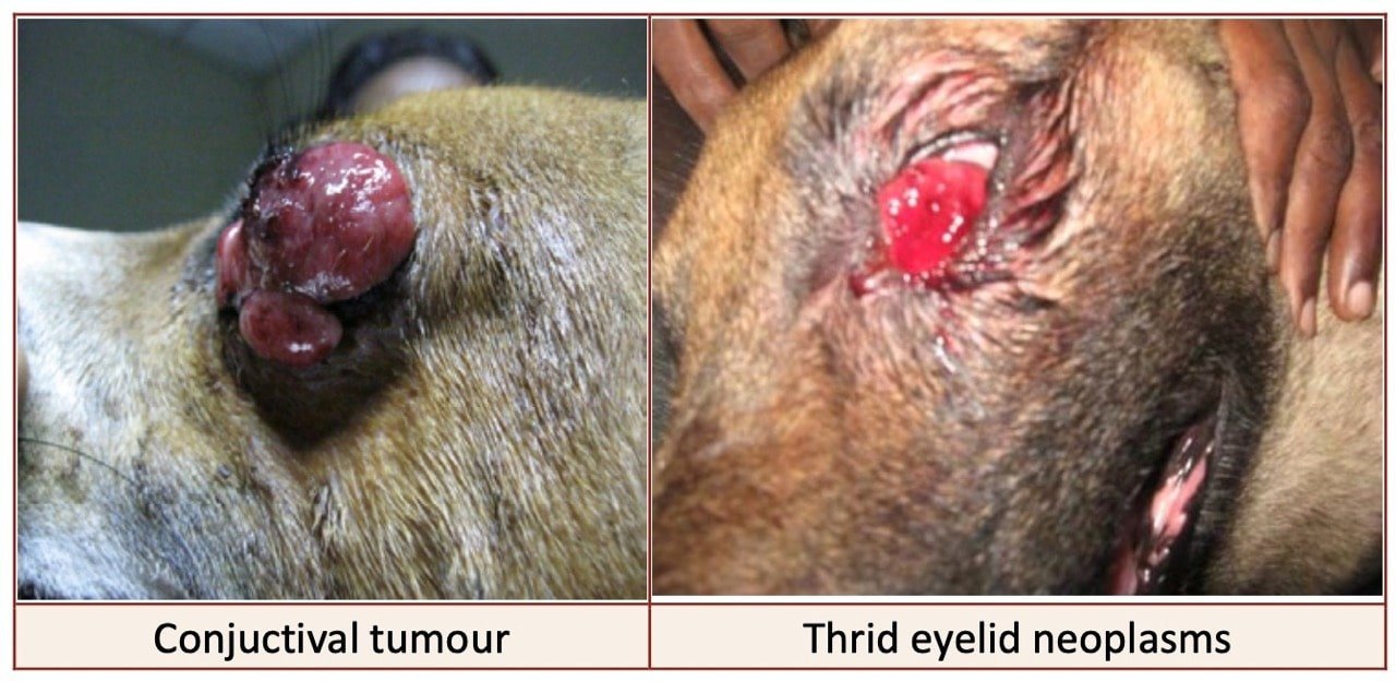
Neoplasms of the third eyelid
Neoplasms of the third eyelid very rare- adenomas, adenocarcinomas and squamous cell carcinomas are reported.

Prolapsed nictitans gland
Prolapsed nictitans gland (prolapse of third eyelid) also known as Cherry eye.
Limbus melanoma
Melanomas may invade the cornea secondarily. These tumors are usually pigmented, occasionally nonpigmented.
The dorsolateral quadrant is usually the site of origination.
Limbal melanomas occur in 2 age groups of dogs.
In the younger group of 2 – 4 years of age, the tumors were invasive.
In the adult dogs 8 – 11 years of age, the tumors were stationary.
Primary limbal melanomas must be differentiated from external extension of intraocular melanomas.
Treatment
Full thickness corneoscleral grafts are recommended to maintain a functional eye in younger dogs with progressive limbal melanomas.
Grafts of nictitating membrane cartilage with overlying conjunctiva have been used to replace corneal and scleral defects after removal of limbal melanoma.
In aged dogs with non progressive limbal masses, periodic surveillance appears to be adequate.
Naso lacrimal duct occlusion
Topical ophthalmic application of fluorescein dye and observation for its appearance at the nares confirms patency of the nasolacrimal duct on that side and is referred to as the Jones or fluorescein passage test.
The interval required for fluorescein to appear is variable (up to 5 to 10 minutes in some normal dogs).
In some dogs and cats, especially brachycephalic breeds, drainage from the nasolacrimal duct may occur into the posterior nasal cavity, resulting in false- negative result of the Jones test unless the mouth is also examined.
Symptoms
Epiphora unilateral or bilateralIf there is obstruction of the duct, a naso lacrimal flushing after catheterisation is practised.
Nasolacrimal Flush (Catheterization) is indicated for epiphora and dacryocystitis.
Procedure for indwelling nasolacrimal duct catheterization for flushing
A monofilament nylon thread (2/0 with a smooth melted end) is passed via the superior punctum to emerge from the nose. If an obstruction is present in the sac, the duct is threaded from the nasal end, and the thread is manipulated to emerge from the superior punctum.
Fine polyethylene (PE90), polyvinyl, or silicone tubing with a beveled end is passed over the thread. Halsted forceps are clamped behind the tubing, which is pulled from the nasal end by forceps on the thread. In horses, larger tubing is used.
Care is taken as the tubing enters the punctum. Note: The inferior punctum may also be used if threading via this punctum was used. The tubing is pulled down the nasolacrimal duct, past any obstructions.
The tube is sutured in place for 2 to 3 weeks. An Elizabethan collar should be considered to prevent the tubing from being dislodged.