TABLE OF CONTENTS
Surgical affections of testicle and Scrotum
Surgical affections of testicle and Scrotum are Cryptorchidism, Orchitis, Trauma of testicles, Tumors of testicles, Affections of epididymus & tubular parts, Affections of prostate gland, Hydrocele etc.
Surgical affections of testicle and Scrotum in animals are-
- Cryptorchidism
- Orchitis
- Trauma of testicles
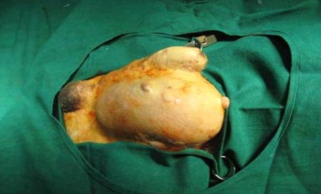
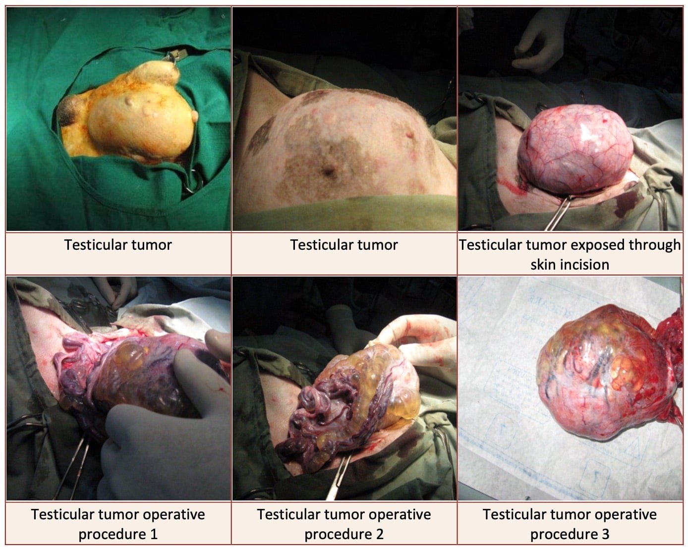
- Tumors of testicles
- Affections of epididymus & tubular parts
- Affections of prostate gland
- Hydrocele
Surgical procedures of testicle and Scrotum in animals are-
Cryptorchidism
In the fetus, the testes are located intra-abdominally near the kidneys. They descend into the scrotum in cats and dogs by about five days after birth. However, normal testicular descend may take six months to complete in some animals.
Failure of the embryonic development of the testicles or their descent into the scrotum can result in congenital abnormalities like anorchism, monorchism, testicular hypoplasia or cryptorchidism.
Anorchism is the congenital absence of both testicles. It is rare in companion animals. Monorchism is the congenital absence of one testicle, the left testicle being usually absent.
Anorchism and monorchism can be diagnosed by careful palpation of the scrotum, inguinal region and the abdomen (intra-abdominal testes are usually palpable only when they are larger than normal). Ultrasonography, laparoscopy or exploratory laparotomy may be required for confirmation.
The conditions are usually asymptomatic except for the failure of development of secondary sexual characters in cases of anorchism and need not be treated.
Testicular hypoplasia may affect one or both testicles. The affected testicles may be located within the scrotum, will be very small, normal or soft in consistency and difficult to palpate.
Usually animals which are bilaterally affected will be sterile. However, the testicular hormones may be produced. Some of the affected animals may show feminization and orchiectomy may have to be performed.
Cryptorchidism is the failure of one or both testicles to descent into the scrotum from the abdominal cavity.
This is the most common congenital condition affecting the testes. Unilateral cryptorchidism is more common and the right testicle is mostly affected.
The ectopic testes may be located in the prescrotal region, inguinal canal or within the abdominal cavity, the latter being more common. The condition may be diagnosed by careful palpation of the prescrotal region, inguinal canal and the abdominal cavity (normal sized intra- abdominal testicles are difficult to palpate). Ultrasonography, laparoscopy and exploratory laparotomy may be required for the diagnosis of intra-abdominal cases of cryptorchidism.
In bilateral cases of cryptorchidism the animal will be sterile as the germinal cells of the testes undergo degeneration in the raised ambient temperature in the ectopic location. However, the endocrine function remains normal and even in cases of bilateral involvement the secondary sexual characters are normal. But, feminisation may be seen in cases where the ectopic testicle/testicles have developed Sertoli cell tumour as intra-abdominal ectopic testicles have a high tendency to develop neoplasms especially Sertoli cell tumour and seminoma. The intra- abdominal ectopic testicles also are prone to suffer from torsion as they are more freely movable.
Treatment involves orchiectomy. Bilateral orchiectomy is preferred even in unilateral involvement to prevent the onward transmission of genes responsible for the condition. The pre-scrotal or inguinal testes are removed through an incision placed on the skin directly over the ectopic testicle. Intra-abdominal testicles are removed through a ventral median laparotomy incision.
Orchitis
Orchitis is the inflammation of the testis, may result from infection of the testicular tissue. The usual route of infection is through the vas deferens from an infected urethra, prostate or urinary bladder. The infection may also reach the testis by a haematogenous route or via a penetrating injury through the scrotal skin.
The condition may be unilateral or bilateral and may be acute in onset or chronic. In acute cases the animal may show pain, tenseness and scrotal oedema. Systemic signs of infection like leukocytosis, fever, anorexia and listlessness may also be seen. The testis appears enlarged and later may get adhered to its tunics. In chronic cases, abscesses may develop which may drain through tracts onto the skin.
Acute cases may be treated using appropriate antibiotics, anti-inflammatory (NSAIDs) or analgesic agents and cold application. In cases of accumulated pus, incisional drainage will be useful in hastening the healing process. Cases that do not respond to conservative measures and are severe or chronic may be treated by orchiectomy.
Trauma of testicles
Trauma of the testicles may result following fights, accidents or attack by man. However, the condition has a low incidence considering the relatively exposed nature of the organs. The affected animal may have swelling of the testis, signs of local pain and even lameness of the hind limbs. Scrotal swelling, bruising and haematoma may be seen in more severe cases.
The tunica albugenia may be ruptured and the testicular tissue may protrude through to variable degrees. Damage to testicular tissue, epididymus and spermatic cord may result in life threatening haemorrhage. Damage to the testicular tissue may lead to temporary or permanent infertility, spermatic granuloma formation due to the antigenic nature of the sperms or immune mediated orchitis.
Mild cases may be treated using local cold application, systemic anti-inflammatory or analgesic agents and antibiotics when possibility for infection is suspected. In cases of severe trauma it is recommended to surgically open the scrotal sac and explore to assess the degree of damage to the testicle.
Any bleeding present should be arrested appropriately. In case of rupture of tunica albugenia with resultant protrusion of testicular tissue, the protruding tissue should be excised and the tunica albugenia sutured with synthetic absorbable suture material. After closure of the skin incision a course of antibiotic should be administered. In cases of extreme irreparable cases of testicular trauma or unresponsive immune-mediated orchitis, orchiectomy may be the treatment of choice.
Tumors of testicles

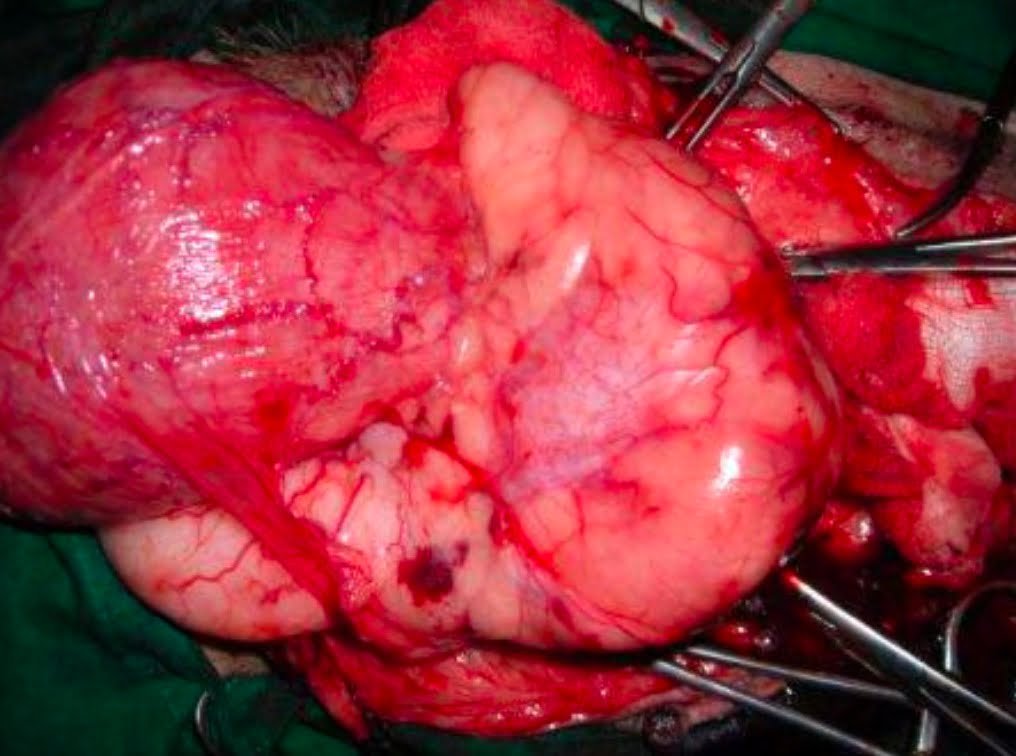
Tumours of the testicle are common in old dogs. The most common are interstitial cell tumours, seminomas and Sertoli cell tumours. Signs include increase in size and firmness of the testicle/testicles, nodular induration on palpation, pain and signs of feminization in cases of Sertoli cell tumours.
The condition should be differentiated from other conditions that can cause an enlargement of the testis or the scrotum like orchitis, torsion of the spermatic cord, testicular or associated tissue trauma, epididymitis, spermatocele, scrotal neoplasms and scrotal hernia. Diagnosis may be confirmed by FNAB or excisional biopsy. Orchiectomy is the treatment of choice.

Affections of epididymus & tubular parts
Conditions affecting the epididymus and the vas deferens can affect the functioning of the genital system. The tubes may suffer from congenital aplasia or occlusion secondary to inflammation/trauma.
Obstruction to the flow of sperm through these channels will lead to the formation of spermatoceles or spermatic granulomas, pain and can cause infertility when bilaterally involved.
Cases of epididymitis may be treated along routine lines but in cases of permanent obstruction orchiectomy may be performed.
Tumours of the epididymus or vas deferens may have to be treated by surgical removal of the affected part and also orchiectomy on the affected side. Bilateral orchiectomy may be performed if further breeding of the animal is not desired.
Conditions affecting the urethra like urethritis and urethral tumours cause signs primarily associated with urine outflow obstruction rather than genital involvement and should be treated appropriately.
Affections of prostate gland
Dogs commonly suffer from prostatic diseases. Male dogs showing tenesmus, dysuria, anuria, pyuria, haematuria, caudal abdominal pain and difficulty in walking with the hindlimbs should be examined for prostatic involvement.
Prostatic diseases are rare in cats. Diagnosis of prostatic diseases may be made from history and clinical signs, per rectal digital palpation of the prostate, plain and contrast radiography, ultrasonography, laparoscopy, biopsy and laboratory evaluation of blood, urine and ejaculate.
Benign prostatic hyperplasia is the most common prostatic disease affecting dogs. It is a normal old age related condition in which the prostate gets enlarged and the enlargement of the gland is testosterone dependant. Constipation, tenesmus, bloody urethral discharge or retention of urine may be seen. Dyschezia is more characteristic than dysuria due to the physical obstruction caused by the enlarged prostate to the expansion of the rectum in the pelvis. Prolonged straining to pass feces may lead to weakening of the pelvic diaphragm and subsequent perineal hernia. Digital palpation per rectum reveals a uniformly enlarged non-painful prostate with a normal spongy consistency. Haemogram and biochemical parameters are usually normal. Bacterial cultures of urine, prostatic fluid and ejaculate are negative. Biopsy may be required for confirmation. However, the latter is reserved for cases that do not respond to treatment.
The recommended treatment for benign prostatic hyperplasia is bilateral orchiectomy. Once the stimulation to the prostatic cells by testosterone is removed, permanent involution of the prostate and clinical relief is obtained in 2 to 3 weeks. In cases where castration is not desired oestrogenic preparations may be used. However, they have the potential to cause feminization and loss of fertility. In valuable animals where it is desirable to retain the fertility, drugs like finasteride may be administered orally. However, the condition may return when the drug is stopped.

Prostatitis and prostatic abscess are not rare findings in dogs. The close proximity of the prostate to the urethra which normally has resident bacteria predisposes it to infection. The condition may be acute or chronic. Clinical signs in acute cases include pyrexia, lethargy, anorexia, urine retention, constipation, purulent urethral discharge, signs of caudal abdominal pain and hind limb gait abnormality. Systemic signs of sepsis may be seen. Palpation of the gland reveals it to be asymmetrically swollen, painful and fluctuant when abscesses are present. Application of pressure on the fluctuating swelling may cause drainage of pus from the urethra. In cases where the abscesses have ruptured signs of peritonitis and septic shock may develop. Urine may be collected and evaluated revealing haematuria and pyuria. Culture of urine and prostatic fluid obtained by catheterization or fine needle aspiration reveals bacteria. Plain and contrast radiography, ultrasonography and laparoscopy may further help in diagnosis.
Treatment
Treatment involves the use of appropriate antibiotics, castration to reduce the size and activity of the prostate, drainage of abscesses, omentalization, marsupialization and partial or complete prostatectomy.
Prostatic and paraprostatic cysts may result from the increased production of prostatic fluid or a structural or functional obstruction to the outflow mechanism. The accumulated secretions may get secondarily infected and form abscesses. Clinical signs may be produced due to the physical obstruction caused by the enlarged cysts as in prostatic abscesses except for the signs related with infection and sepsis. Diagnosis is also made by the techniques described earlier. Culture of the prostatic secretions reveals no bacteria except in cases with secondary bacterial infection.
Surgical treatment is aimed at drainage, removal or debulking of the affected prostatic tissue and omentalization of the remnants. Castration is also recommended.
Prostatic tumours typically affect old dogs and can be prevented by castration. Though adenocarcinoma and transitional cell carcinoma are most common in dogs other types have also been reported. Clinical signs are produced by the physical obstruction to the urinary and fecal outflow. Also, other signs of neoplasia like cachexia, anorexia and pain will also be pronounced.
Metastasis to adjacent and distant organs also produces related symptoms. Rectal or abdominal palpation reveals a painful, firm, irregular and nodular prostate which may or may not be adherent to the surrounding structures. Lymphadenopathy may be palpable or may be ultrasonographically visualized. Biopsy may be performed for differentiation of the condition from other conditions that cause an enlargement in the size of the prostate.
Treatment by prostatectomy may be performed before the tumour has started metastasizing. Advanced cases have poor prognosis.
Trauma of prostate may occur because of trauma to the pelvic region resulting in pelvic fractures or penetrating caudal abdominal injuries. Mild cases may be treated by establishing the patency of the urethra by catheterization and allowing the damaged gland to heal by second intention.
In cases where catheterization cannot establish patency of the urethra an exploratory laparotomy may be performed and the damaged prostate may be repaired by suturing the capsule. Partial or excisional prostatectomy may be performed in severe cases of prostatic trauma.
Hydrocele
Hydrocele is the accumulation of fluid in the tunica vaginalis, may result from trauma to the testicle or faulty castration technique using Burdizzo castrator in bulls. Surgical treatment involves orchiectomy on the affected side by the open-covered method. In cases of bilateral involvement, bilateral orchiectomy and scrotal ablation may be performed.
Orchiectomy
Orchiectomy is a common surgical procedure in companion animals performed for management, prophylactic and therapeutic purposes.
Bilateral orchiectomy renders the male animal benign and easier to manage, prevents roaming especially in search of females in heat, reduces injuries due to fighting and prevents development of prostatic hyperplasia.
Orchiectomy is also performed to treat prostatic diseases, perineal hernia and irreparable injuries/neoplasms affecting the testis.
In dogs, the surgery is usually performed by the open method by a prescrotal approach under general anaesthesia. After controlling the animal on dorsal recumbency and preparation of the prescrotal and scrotal skin, a midline incision is placed on the prescrotal skin after tensing one of the testicles under the skin.
The incision extends through the skin, subcutaneous tissue and the tunica vaginalis. The testis is squeezed out and the attachment of the epididymus to the tunica vaginalis is separated bluntly by traction or transected. The vascular and the avascular bundles of the spermatic cord are separated.
The vascular bundle is ligated using No. 1-0 catgut and transfixed. The ends of the suture material may be used for ligating the avascular bundle also. The spermatic cord is transected distal to the ligation and the stump returned into the tunica vaginalis. The other testicle may be removed through the same skin incision by incising the scrotal septum after tensing the testicle against it.
The procedure is repeated to remove the second testicle. Subcutaneous sutures may or may not be placed using No. 4-0 absorbable suture material and the skin incision can be closed using No. 3-0 or 4-0 nylon.
In cats, orchiectomy is performed by placing separate longitudinal incisions on the scrotal skin over each testicle.
The spermatic vessels may be ligated as in the dog or the vascular and avascular components of the spermatic cord may be used for arresting bleeding by applying two square knots with them. The scrotal skin incision may be left without suturing.
Castration
Castration in farm animals
Cattle, sheep and goats are usually castrated by the closed method using Burdizzo castrator. After controlling the animal in lateral recumbency with appropriate restraint by tying up the fore and hind legs together, the spermatic cord on one side is identified.
The spermatic cord is kept tensed against the scrotal skin and trapped within the jaws of the castrator. The arms of the castrator are approximated thereby crushing the spermatic cord.
The castrator is removed and the procedure is repeated on the other side taking care that the crush lines on the scrotal skin on either side do not meet to avoid sloughing of the scrotal skin distal to the crush lines.
The crushing of the spermatic cord may be repeated at two levels on each side if desired.
Castration in horses
Castration of horses is performed under general anaesthesia by the open-covered method. After restraning the animal on lateral recumbency and aseptic preparation of the scrotal and surrounding skin, a longitudinal incision is made on the scrotal skin over one testicle. The tunica vaginalis is incised and the testicle exteriorized.
The vascular and avascular bundles are doubly ligated using heavy catgut. The spermatic cord is crushed and transected distal to the ligation using an emasculator and the stump returned as high as possible in the external inguinal ring.
The tunica vaginalis is ligated as high as possible and transfixed and the part distal to the ligation transected and removed. Alternatively, the tunica vaginalis may be transected close to the level of the external inguinal ring and the edges apposed and sutured.
The scrotal sac and the ventral aspect of the inguinal canal may be packed with sterile gauze which can be kept in place for two days to stimulate inflammation and early closure of the inguinal canal to prevent chances of inguinal herniation.
The procedure is repeated on the other side to remove the remaining testicle. In addition to a post-operative course of antibiotic an appropriate dose of tetanus toxoid should also be administered.
Vasectomy
Vasectomy inhibits male fertility but maintains behavioural pattern.
Procedure
- Make a 1 to 2 cm incision over the spermatic cord between the scrotum and inguinal ring
- Locate spermatic cord, incise vaginal tunic
- Isolate the ductus deferens by blunt dissection
- Double ligate ductus deferens and resect a 0.5cm section of ductus between ligatures
- Repeat the same on the contralateral spermatic cord.
- Appose subcutis and skin
- Vasectomy – reduces hormone associated diseases.
- But not roaming, aggression and urine marking
- Therefore it is not much recommended in canines
- Androgens are continually produced within one week the animal becomes azoospermic following vas occlusion
- But spermatozoa may persist in ejaculation for 3 weeks(canines), 7 weeks in felines after vasectomy
Granuloma, scrotal swelling, incisional problems are may be some complications of Vasectomy.