TABLE OF CONTENTS
Surgical affections of Salivary glands
Surgical affections of Salivary glands in animals are open wounds, salivary fistula, salivary Mucocele, Ranula (Honey cyst), foreign bodies in the salivary ducts, salivary calculi, tumors and parotid abscess.
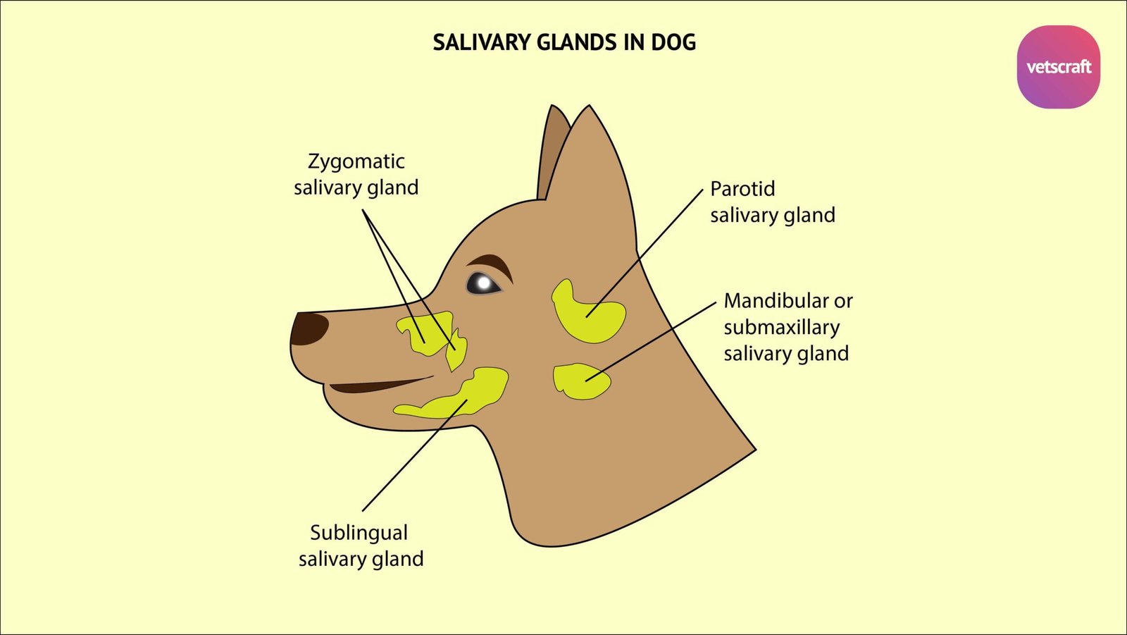
Major salivary glands presents in animals are-

- Parotid salivary glands
- Mandibular or submaxillary salivary glands
- Sublingual salivary glands
- Zygomatic salivary glands
Surgical affections of salivary glands in animals are-
- Open wounds
- Stenson’s duct wound
- Salivary fistula
- Foreign bodies in the salivary ducts
- Salivary calculi
- Tumors
- Parotid abscess
- Salivary Mucoceles
- Ranula (Honey cyst)
Open wounds of salivary glands
If any salivary gland is wounded, there is an escape of saliva through the wound.
Treatment of open wounds
Arrest the haemorrhage by appropriate measure. If some of the large vessels are severed, they must be promptly secured by artery forceps and then ligate.
If the vessels cannot be secured by artery forceps, the vessel should be isolated at some distance from it and then the ligature is applied.
Ligation of the carotid may not stop, persistent haemorrhage from the internal carotid noticed, it can be anastomosed with the corresponding artery on the other side. Suture the wound and apply bandage.
Stenson’s duct wound
Wounds of the duct may be transverse, longitudinal or oblique and partial or complete.
During feeding, there is copious discharge of saliva from the wound. It is more difficult to obtain healing of the lesion here than in the gland.
Treatment of Stenson’s duct wound
For promoting healing by first intension, perforate the cheek at the level of the wound, when the latter is near the mouth, to provide another passage for the saliva.
Suture the cutaneous wound and bandaged with povidone-iodine. The patient should not be given any solid food for 24 hrs.
Salivary fistula
Salivary fistula in animals may be due to a wound of the parotid, or submaxillary, salivary gland or of Stenson’s duct or Wharton’s duct.
Etiology of Salivary fistula
Salivary fistula may be caused by an open wound or an abscess involving the canal.
Treatment of Salivary fistula
Salivary fistula occurs due to a recent incised wound of the gland or duct, endeavour to get healing by first intension. If the fistula has been in existence for some time, cauterisation of its edges or freshening is done. After that, suture should be done.
Application of silver nitrate or pure nitric acid with a glass rod or of the hot iron, or freshening the lips of the wound and inserting a purse- string suture is often successful. After that there is no obstruction to the flow of saliva.
If the above methods fail, Destruction of the function gland may be adopted:
- Injection of irritants
- Ligation of Stenson’s duct
- Clearing the lumen of the distal portion of the duct
- Making an artificial opening into the mouth
Foreign bodies in the salivary ducts
Small particles may enter into salivary glands and set up inflammation. Foreign bodies rarely causes irritation and form a calculus.
Treatment of Foreign bodies in the salivary ducts
To remove the foreign body, give pressure on the course of the duct. Otherwise, oral orifice of the canal may be incised or Wharton’s duct may be incised.
Apply antiseptic mouth lotion for a few days.
Salivary calculi
Salivary calculi in animals usually occur in Stenson’s duct and rarely in Wharton’s duct.
The sizes of calculi weighing individually 7-12 ounces have been recorded (mention in grams).
An isolated calculus is oval; smooth and yellowish grey in colour. Composition of deposit 80-90% calcium carbonate and 9-10% organic matter.
Etiology of Salivary calculi
Small particles with bacteria carrying into the duct, which cause fermentation in the saliva. Consequent deposition of lime on the foreign body.
Treatment of Salivary calculi
First force the calculus into the mouth and then remove it. If it bulges into the oral cavity, incise the mucus membrane covering the calculus and take it away.
Suture the wounds in the duct and skin separately, using a very fine needle.
Tumors of salivary glands
The common tumors in parotid salivary glands are melanomata, which are found chiefly in grey horses.
If the tumor is benign and circumscribed surgical excision is carried out. If it is malignant or diffuse, it is better not to interfere.
Parotid abscess
Parotid abscess is a painful inflammatory swelling. Strangles in the horse and tuberculosis in the ox may lead to abscess in subparotid region.
Parotid abscess may burst in the course of 8-14 days.
Treatment of Parotid abscess
Open the abscess and drain the content. It may be possible to open the abscess bluntly after incising the skin.
Use suitable antiseptic dressing and systemic antibiotic.
Salivary Mucoceles
Salivary Mucoceles is the collection and accumulation of saliva due to blockage of the duct and is surrounded by granulation tissue.

Symptoms of Salivary Mucoceles is presence of a swelling in the site subcutaneously.
Treatment of Salivary Mucoceles
Salivary Mucoceles is treated by establishment of the patency of the duct:
- Mandibular and sublingual salivary gland excision in Mucocele
- Zygomatic gland excision in zygomatic Mucocele.
- Parotid gland excision
- Destruction of gland
Ranula (Honey cyst)
Ranula or Honey cyst is a retention cyst due to obstruction of mucus glands or duct of sublingual salivary gland. Occurs under the tongue close to the frenulum linguae. Disturbed prehension and excessive salivation, Interferes with mastication, swallowing.
Treatment of Ranula (Honey cyst)
Excision of cyst or opening and painting gland with Povidone Iodine solution.