TABLE OF CONTENTS
Surgical affections of ovary and uterus
Surgical affections of ovary and uterus are Metrorrhagia, Metritis, Pyometra, Uterine torsion, Uterine rupture, Vaginal Fibroma, Ovarian cysts, Ovarian tumors, Vaginal prolapse or hyperplasia etc.
Surgical affections of ovary and uterus in animals are-
- Atresia or occlusion of the Os uteri
- Wounds of the Uterus
- Metrorrhagia
- Metritis
- Pyometra
- Uterine torsion
- Uterine prolapse
- Uterine rupture
- Vaginal Fibroma
- Ovarian cysts
- Ovarian tumors
- Vaginal prolapse or hyperplasia
Surgical procedures of ovary and uterus in animals are-
Atresia or occlusion of the Os uteri
This Atresia or occlusion of the Os uteri condition may be due to a neoplasm or cicatricial contraction.
It renders impregnation difficult or impossible.
Treatment
When the opening is not completely obliterated, it may be dilated with the fingers or special dilators.
Wounds of the Uterus
Wounds of the Uterus may be confined to its mucous membrane or extend more deeply and perforate abdominal cavity.
Gravid uterus may be rupfused by violent impact of the abdominal wall against a fixed object.
Treatment
Rupture during gestation, all that can be done is to treat for internal haemorrhage.
Non perforating wounds inflicted at the time of parturition are treated by antiseptic irrigation and antiseptic cpessaries.
When the organ is perforalted, these is no effective treatment for the condition.
The administration of sedative medicine to allay straining may help to bring about spontaneous recovery.
Metrorrhagia
Haemorrhage from the uterus is usually the result of a round inflicted during parturition.
Treatment
- Cold douches over the loins
- Injections of cold or very hot water into the uterus
- Packing the uterus and vagina with sterilized cloths
- Hypodermic injection of adrenaline is more effective
- Packing material should be removed after 24 hrs
- Uterus should be irrigated with suitable antiseptic solution
Metritis
Metritis is the inflammation of uterus due to presence of pathogenic bacteria, local inflammation, febrile disturbance, offensive mucopurulent discharge from the vagina.
Treatment
- Repeated irrigation by antiseptic solutions, antiseptic pessaries
- Administering suitable medicine internally, including pericillin
- In chronic cases autogenous vaccine is indicated
Pyometra
Pus formation in the uterus is known as Pyometra. It is opened and closed pyometra.


Treatment
Medical treatment help in some extend only, best go for OHE.
Uterine torsion
Uterine torsion condition is uncommon in dogs and cats. The gravid or non-gravid uterus can rotate clockwise or counter-clockwise from 90° to more than 200°.
Etiology
Jumping during or running late in pregnancy, active fetal movements, premature uterine contraction
Treatment
Ovariohysterectomy and c-section if viable foetuses are present.
Uterine prolapse
Uterine prolapse is the prolapse of the uterus through the vaginal canal due to the weakening of its support structures or other underlying conditions.
Etiology
Excessive relaxation and stretching of pelvic musculature, uterine atony due to metritis, incomplete separation of placental membranes, severe tenesmus, post partum contractions intensified by oxytocin release during lactation.
Treatment
If animal is in good condition manual reduction maybe attempted. Sterile gauze soaked in warm sterile saline placed around the uterus. General or epidural anaesthesia is usually necessary.
Extensive uterine devitalization needs ovariohysterecomy after reduction of prolapse. If reduction is not possible the uterus is amputated and stump reduced.
Uterine rupture
Rupture of gravid uterus rare occurrence and can occur during parturition or after severe trauma.
Foetuses expelled into the abdominal cavity may die immediately or be reabsorbed or remain intact, causing peritonitis. If foetal circulation remains intact foetuses may live to term.
Clinical signs may be abdominal enlargement or a palpable abdominal mass. If the tumor obstructs the lumen mucometra or hydrometra may develop.
Treatment
Ovariohysterectomy uncommon in dogs and cats. One or both the horns may prolapse during prolonged labour or upto 48 hours after parturition when the cervix is extremely dilated.
Vaginal Fibroma

Ovarian cysts
Ovarian cysts are Follicular cyst, Lutein cysts and Paraovarian cyst.
Follicular cyst develop from graffian follicles Clinical signs include prolonged estrus with bloody vaginal discharge, cystic mammary hyperplasia,
Lutein cysts develop from corpus luteum after ovulation, maybe associated with cystic endometrial hyperplasia or pyometra. Mostly asymptomatic and found during routine ovariohysterectomy or laparotomy.
Paraovarian cyst originate either from remnants of mesonephric or paramesonephric ducts and tubules. More common in dogs than in cats. Located between ovaries and ovaries and uterine horns. No clinical signs and found incidentally.
Ovarian tumors
Ovarian tumors are large tumors palpable in the cranial right or left abdomen. Surgical treatment includes ovariohysterectomy.
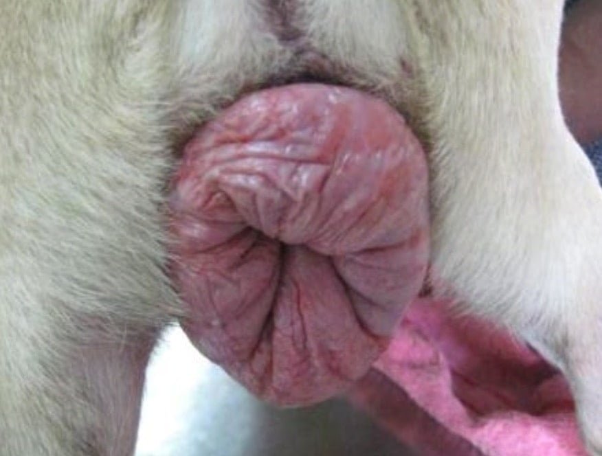
Vaginal prolapse or hyperplasia
Vaginal prolapse or hyperplasia occurs as a result of edematous enlargement of vaginal tissue during estrus or proestrous.

Vaginal prolapse occurs as a 360 degree involvement of the protrusion of the mucosa where as hyperplasia arise from a stalk of mucosa from the vaginal floor. weakness of the vaginal connective tissue results in edema and prolapse through the vulva.
It occurs in young bitches 2 years or younger and is extremely rare in cats.
Differential diagnosis
The most common types of vulval vaginal tumors are fibroleiomyoma, sqaumous cell carcinoma and transmissible venereal tumor.
Treatment
- If the protrusion is small the prolapse will resolve once the effects of estrogen diminishes.
- For this GnRH can be given at the dose rate of 50 microgram / 40 lb bodyweight. In TVT Vincristine can be administered at the dose rate of 0.025 mg/kg up to 1 mg IV weekly for 3-6 weeks
Surgical treatment
- OHE is recommended to prevent injury to the evereted mucosa
- Manual reduction after episiotomy and suturing the vulval lips till edematous stage resorbs.
- Resection of the protruding mass with OHE is recommended if the tissue is severly damaged.
- Resection of the protruding mass without OHE may require hysteropexy, cystopexy or colopexy, but this is not practiced in TVT cases.
Ovariohysterectomy (OHE)
Ovariohysterectomy (OHE) is the surgical removal of the uterus and ovaries under general anaesthesia. This procedure is typically performed around or prior to six months, but can be performed on dogs of any age.
The procedure may be elective, or a treatment for a disease process.
Reasons for performing the surgery
- Vastly decreased chance for development of mammary cancer
- 200 times less likely if ovariohysterectomy performed before the first estrus
- Eliminates chance of developing a pyometra or uterine infection
- Eradicates unwanted estrous behavior and associated bleeding
- Eliminates unwanted pregnancies and risks of dystocia (difficult birth)
Anaesthesia
- Premedicate with atropine, followed 10 minutes by xylazine @1 mg/kg body weight. Induce the anaesthesia with ketamine @ 10 mg/kg body weight and diazepam 0.3 mg/kg body weight.
- Maintain anaesthesia with same ketamine and diazepam or propofol @ 3-5 mg/kg body weight.
Preparation of the animal
- Position the animal in dorsal recumbency or left lateral recumbency
- Prepare the area aseptically
Procedure
- The surgical incision is usually made along the ventral abdomen, but flank approaches have been reported
- Separate the subcutaneous tissues and facia. Incised linea alba. The ovary is identified and surgical clamps are applied to the ovarian blood vessels.
- The vessels are then ligated (tied with sutures) to prevent bleeding and the pedicle is replaced into the body. This procedure is repeated for the other side
- The uterus and its blood vessels are ligated just above the cervix

- The uterus and ovaries are removed from the abdomen. The abdomen is sutured closed in three layers: the abdominal wall, the subcutaneous tissue (tissue underneath the skin) and the skin itself.
Complications
Ovariohysterectomy can lead to mild complications such as incisional bruising, swelling and infection. More serious complications such as hemorrhage and urinary obstruction are rare but can be life-threatening.
Ovariohysterectomy can be more difficult in larger or obese animals and may be associated with more complications.
Postoperative care
After care includes house rest, with no running, jumping or rough play for two weeks following surgery. Pain medications are often prescribed for several days following surgery.
An Elizabethan collar may be necessary to prevent licking of the surgical wound. Further treatments may be necessary following ovariohysterectomy for treatment of pyometra or other disease.
Prognosis
The prognosis is excellent for routine ovariohysterectomy. Prognosis is good following ovariohysterectomy for pyometra and dystocia.
Spaying
Removal of the ovary is known as spaying.
Indications
- Prevent breeding nuisance
- Prevent development of pyometra, mammary tumor
Age to perform
Above 6 months of age in case of dogs.
Anaesthesia
- Premedicate with atropine, followed 10 minutes by xylazine @1 mg/kg body weight.
- Induce the anaesthesia with ketamine @ 10 mg/kg body weight and diazepam 0.3 mg/kg body weight.
- Maintain anaesthesia with same ketamine and diazepam or propofol @ 3-5 mg/kg body weight.
Preparation of the animal
Position the animal in dorsal recumbency or left lateral recumbency. Prepare the area aseptically.
Sites
- From a point a little behind the umbilicus backwards along the midline over a length of 3 -5 inches.
- 2.1 – 1 1⁄2 inches incision on either flank, parallel to the last rib, below the lumbar transverse processes, at the level of the posterior lobe of the kidneys.
- The incision may be 1⁄2 inch behind the last rib on the right flank and about 1 inch behind on the left flank.
Technique
- Perform laparotomy
- The ovary with its bursa is held with fingers
- A ligature is applied anterior to the ovary and another one behind it, around the respective vascular connections
- The ovarian bursa is opened and the ovary is removed learning the bursa.
- The other ovary also is removed in a similar manner
- The abdomen is sutured closed in three layers: the abdominal wall, the subcutaneous tissue (tissue underneath the skin) and the skin itself
Postoperative care
- Aftercare includes house rest, with no running, jumping or rough play for two weeks following surgery
- Pain medications are often prescribed for several days following surgery
- An Elizabethan collar may be necessary to prevent licking of the surgical wound