TABLE OF CONTENTS
Surgical affections of Oesophagus
Surgical affections of Oesophagus in animals are Choke of oesophagus, Foreign body, Esophageal Strictures, Vascular ring anomalies, Periesophageal Masses, Megaesophagus, Esophageal Diverticulum, Esophageal perforation and laceration, Esophageal fistula etc.
Surgical affections of Oesophagus are-
- Choke of oesophagus
- Foreign body
- Esophageal Strictures
- Vascular ring anomalies
- Periesophageal Masses
- Megaesophagus
- Esophageal Diverticulum
- Esophageal perforation and laceration
- Esophageal fistula
Choke of oesophagus
Choke of oesophagus is obstruction of the oesophagus in animals. It is most common in bovines as they have the habit of indiscriminate feeding and thus picking up the foreign bodies especially during pregnancy.
Seat of obstruction
- All animals: Just behind the pharynx
- Horse: Inferior third of oesophagus, being normally constricted
- Ox & dog: Lower part of cervical region due to its compression between the thoracic inlet and the first rib
Chocking objects
- Horse: Carrot, turnip, potato, piece of wood, extracted tooth and broken balling gun swallowed accidentally
- Ox: Turnip, potato, apple, palm or mango kernel
- Dog and Cat: Bone or cartilage, (fixation is due to sharp points) swallowing of foreign bodies while playing
Symptoms
- Cessation of feeding
- Makes frequent gulping movements
- Frequent attempts at vomiting (arches the neck, bring the muzzle towards the chest)
- Salivation when the obstruction in near the pharynx
- Tympany or bloat in large animals
- swelling or bulging of the oesophagus at the cervical region in cervical obstruction
Treatment
Medical management is proceed by emetic agents, Cervical oesophagotomy is a surgical management is required when medical management of choke is not work.
Foreign body
Foreign body also leads to esophageal obstruction in small animals.
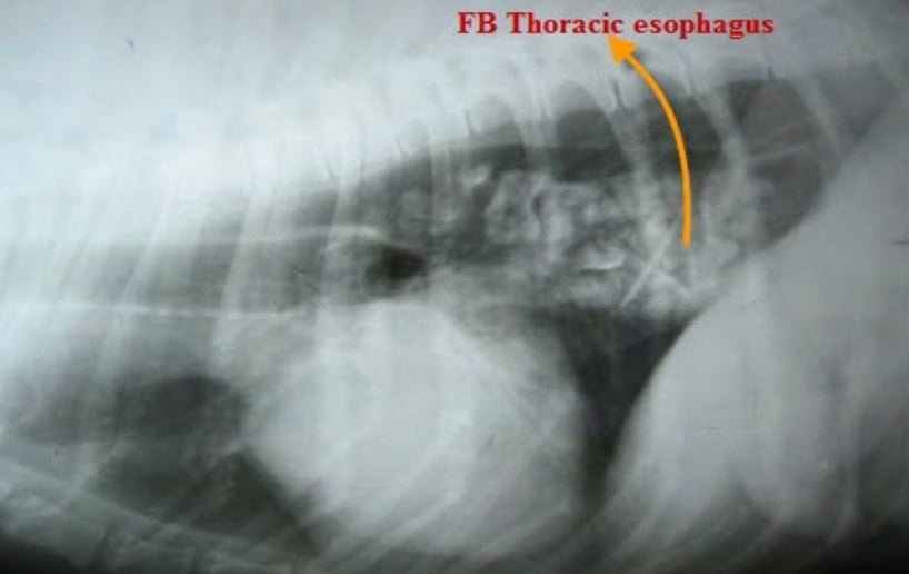
Foreign body in Oesophagus in a dog
The most common cause for esophageal obstruction is ingestion of foreign bodies. Various objects may lodge and produce partial or complete obstruction in oesophagus. The most common foreign body is bones.
Others include needle, wooden sticks, rubber toys, plastics and coins. Cats are more predisposed to ingesting fishhooks and needles. The ingested foreign bodies become lodged in the cervical constriction, bronchoaortic constriction, diaphragmatic constriction and thoracic inlet.
Most of the foreign bodies produce acute clinical signs because of either complete obstruction or severe, painful, partial obstruction.
Longer the duration of large, sharp foreign body, obstruction is more prone to serious complications.
Surgical management is indicated when the conservative treatment fails. Thoracic esophagotomy is more complicated than cervical esophagotomy. If the object is located caudal to the base of the heart, the foreign body can be removed via an abdominal gastrotomy. If possible the foreign body may be crushed into small pieces to help easy removal. Alternative method is to perform the gastrotomy via a thoracic, transdiaphragmatic approach. Left sided 8 th intercostal thoracotomy is performed, the lungs are packed off, the diaphragm is incised to expose the greater curvature of stomach. Spillage of the gastric content in the thoracic cavity is the important complications in thoracotomy.
Difficulty in swallowing ,regurgitation, gulping, excessive salivation and inappetence (cats) are the acute clinical signs. Chronic signs are Cervical swelling, primary malnutrition and aspiration pneumonia.
Pre operative management of patients with esophageal disorders correct fluid electrolyte and acid base imbalance.
Prophylactic antibiotics for esophagitis an aspiration pneumonia support nutrition.
If the object has perforated and cannot be removed, surgery can be used to cut off the extraluminal foreign objects while the intraluminal foreign body can be recovered with the endoscope. If the foreign body is a bone it may be pushed into the stomach rather than removed by orally. Most of the bones are quickly digested in the gastric acids and excreted within 10 days in the feces. If the signs develop that may indicate the surgical removal of the remaining bones.
Esophageal Strictures
Acquired esophageal stricture caused by any damage to the mucous membrane that produces injury by foreign body, sequelae to previous esophagotomy, external compression of the lumen by the presence of parasitic tumour (spirocerca nodule), Compression of the oesophagus by tumours, abscess (extra esophageal mass ) Congenital esophageal stricture is rare in dogs.
Surgical management is more complicated in this case. If treatment is aimed at resection and anastomosis , surgeon may inadvertently leave behind damaged tissue, which leads to reformation of the stricture.
The resected esophagus length is more, the anastomosis may fail due to tension between anastomosis part. In that situation, patch grafting, muscle interposition graft, circular myotomies, suture line reinforcement or segmental replacement can be use as an alternative techniques.
The best treatment for Esophageal Strictures is mechanical dilation and pharmacological intervention with agents that reduce fibroplasia and collagen cross-linking.
Vascular ring anomalies
Vascular ring anomalies is the most common cause of extraluminal esophageal obstruction in dogs and cats. Due to this kind of chronic partial obstruction, which causes serious complications include proximal dilation, loss of motility in the dilated segment, ulcerative esophagitis, cachexia and aspiration pneumonia.
Vascular ring anomalies are the result of abnormal development of definitive vascular structures derived from embryonic aortic arches.
Clinical signs result from partial or complete entrapment of the esophagus between the base of the heart and the affending vessels. Mechanical obstruction is produced by vascular ring itself but concurrent fibrosis of the underlying esophageal wall develops.
Most common vascular ring anomaly in dog and cats results from persistence of the right fourth aortic arch as the definitive aorta. Stenosis of the esophagus occurs due to ductus arteriosus.
Affected animals are considered normal until weaning. Liquids bypass the esophageal obstruction without difficulty. As an animal ingests solid foods, postparandial regurgitation occurs. Megaesophagus develops early in the disease.
Diagnosis of vascular ring anomalies is based on history, physical examination, radiography and endoscopy. Megaesophagus may be diagnosed on physical examination by observing and palpating a bulge in the ventral cervical and thoracic inlet after swallowing.
Treatment is surgical, requiring division of the appropriate portion of the ring to relieve esophageal stenosis.
Periesophageal Masses
Mechanical obstruction of esophagus may occur secondary to lesions in surrounding tissues. Esophageal dysfunction in cervical region caused by thyroid carcinoma, laryngeal carcinoma, salivary gland neoplasms, squamous cell carcinoma and metastatic tonsillar carcinoma in cervical lymphnodes.
Abscess and granulomas may also cause this. In the thoracic inlet, cranial mediastinum and thoracic cavity other lesions including thymomas, lymphomas, large lung tumors, abscesses, and granulomas may occur.
Clinical signs of partial obstruction includes regurgitation, salivation, discomfort, dysphagia, cough and dyspnea if aspiration occurs. Infiltration through the esophageal wall can be determined by endoscopy preoperatively.
Neuromuscular diseases
Mechanical obstruction of the esophagus and propulsion of ingesta into the stomach results from inherent disorders of esophageal function.
Most of this conditions managed by medically but surgery is necessary in some cases.
Megaesophagus
Megaesophagus can develop cranial to the mechanical obstruction.
Generalised megaesophagus can result from affections of vagus, metabolic diseases like hypo-adrenocorticism, hypothyroidism and immunological diseases like myasthenia gravis, polymyositis, and certain drugs such as anticholinergics, general anaesthetics , and idiopathic disorders. These are managed well by medically and feeding in upright position than surgically.
Other disorders like cricopharyngeal achalasia and gastroesophageal achalasia also occurs.
Esophageal Diverticulum
Focal out pouching of the esophageal wall is called Esophageal diverticulum. This may be congenital or acquired but not common in small animals.
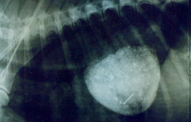
Contrast medium showing Esophageal Diverticulum in a dog
Congenital diverticulum results from inherent weakness of the esophageal wall, failure of the primordial foregut and pulmonary buds to separate or eccentric vacuole formation in the esophagus.
Acquired diverticulum are two types. Depending on their cause and histological appearance they are called as pulsion diverticulum or traction diverticulum.
Pulsion diverticulum
Pulsion diverticulum is an outpouching of mucosa through a defect or tear in the overlying muscularis. This is otherwise called as false diverticulum because not all layers of the oesophagus are represented in the protruding sac. This will develop after focal pathological pressure applied to esophageal wall from within the lumen. It may also result from regional abnormalities in peristalsis in association with obstruction.
The most common site of diverticula is just proximal to diaphragm. Dysphagia, regurgitation, gagging, gulping weight loss and respiratory signs are usual clinical signs. Contrast radiography and endoscopy are effective diagnostic methods.
The diverticula may be large and sometimes multiple and often impacted with ingesta. Small diverticulum may be managed conservatively by diet modification and upright feedings. If the diverticulum is too large, resection of the diverticulum is indicated.
The diverticulum is single and focal, simple excision of the sac with two layer repair of the esophageal wall is sufficient. For large and multiple diverticula, resection and anastomosis or hemicircumferential wall resection and reconstruction is required.
Traction diverticulum
Traction diverticulum is otherwise called as true diverticulum, which is composed of all layers of the esophageal wall.
They termed “traction” because of their presumed pathogenesis, involving the adhesion and contraction of fibrous band to esophageal wall results in outpouching.
The causes are local inflammation outside the esophagus which includes disease processes involving the trachea, lungs, hilar lymph nodes and pericardium.
Esophageal perforation and laceration
Esophageal perforation and laceration may occur from inside or outside the oesophagus. Bite wounds, gunshot wounds lacerations due to vehicle injuries may result in perforation or laceration of oesophagus. Also results from ingestion of sharp foreign bodies with or without signs of obstruction.
Clinical signs depend on the location, extent and duration of the perforation and associated leakage.
The inflammation, hypoxia and necrosis in local tissues may predispose to massive infection. Saliva, ingesta and microorganisms may leak from the esophagus which causes local cellulitis and abscess.
The perforation confirmed with esophagoscopy or contrast esophagography. Conservative management includes antibiotics, with holding of food and water for several days and maintenance of hydration and electrolyte balance.
In leakage, the perforation is exposed and the esophagus repaired primarily. If the wound is unhealthy, and they are debrided and a two-layer closure technique can be used. If the wound is chronically infected, a reinforcing technique is used. Postoperative care includes 3 to 5 days of esophageal rest, using parenteral or gastric alimentation.
Esophageal fistula
Esophageal fistula is an abnormal communication between the esophagus and the trachea, bronchus, lung parenchyma or the skin.
Congenital fistulas occur due to failure in complete separation of the developing foregut and respiratory tracts. Acquired fistulas are more common which arises secondary to trauma.
Esophagobronchial fistulas are more common than esophagotracheal and esophagopulmonary fistulas. In dogs the fistulas most commonly occurs between oesophagus and the right caudal lung lobe. In cats, they are in the accessory lobe and left caudal lung lobe.
Cough induced by ingestion of food or liquids but in some cases chronic signs of pneumonia or lung abscessation may occur. Positive contrast radiography can be used to demonstrate direct communication between esophageal lumen and respiratory tract.
Treatment involves thoracotomy to expose the oesophagus , fistula and affected portion of the respiratory system.