TABLE OF CONTENTS
Surgical affections of Neck
Surgical affections of Neck in animals are Poll Evil, yoke gall, yoke tumor and yoke ulcer, affections of wither like Fistulous withers (supraspinous bursitis) and poll evil etc.
Surgical affections of Neck in animals are –
Poll Evil
Poll Evil is due to necrosis of ligamentum nuchae and dorsal arch of the atlas in horses.
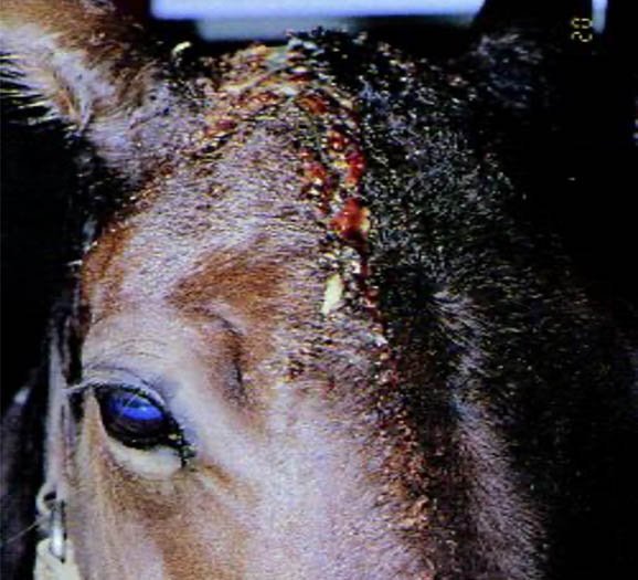
Poll Evil in a horse
Etiology
Injury and infection of the poll leads to Poll Evil.
Symptoms
- Inflammatory swelling, which is very painful and lobulated
- Purulent discharge
- Head is kept abnormally high due to pain
Prognosis
Prognosis is not serious but, very troublesome as it is very difficult to get rid of all the necrotic tissue and provide effective drainage for the pus.
Treatment
Surgical excision under general anaesthesia is required. 5-8 cm long incision in the middle line of poll from in front of occipital crest to a point behind the posterior limit of the lesion.
Procedure
- Incise through skin down to the ligament nuchae.
- Disect it out as far as it is diseased.
- Sever it posteriorly, reflect it outwards, from its insertion into occipital crest.
- Curette the tracks of the ligament and ulcers on the bone.
- Remove all necrotic tissues from its depth.
- Arrest the profuse haemorrhage by plugging with sterile medicated gauze. for 24 hours Thereafter treat it as an open wound (for 6 weeks)
Result
First the animal feels difficulty in grazing but in course of time this inconvenience disappears due to the stretching of the new cicatrix.
Cutaneous horn or cornu cutaneum
Cutaneous horn or cornu cutaneum is a protrusion from the skin consisting of cornified material organized in the shape of a horn.
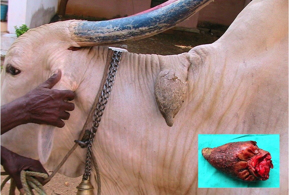
Yoke gall and tumor
The surgical affections of this region are yoke gall, tumour and yoke ulcer. These are seen in the cattle and buffaloes.
Yoke Place
Normally there is no bursa in Yoke Place region, but due to constant pressure of the yoke an acquired subcutaneous bursa develops.
Yoke gall is the localized acute inflammation of the skin and subcutis on the neck due to injury caused by friction (rubbing) of the yoke. It may be a swelling due to separation of layers of skin and subcutis and accumulation of inflammatory exudates there in.
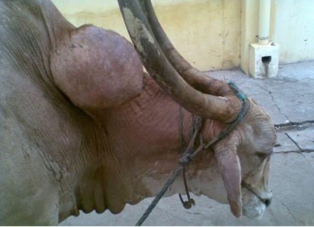
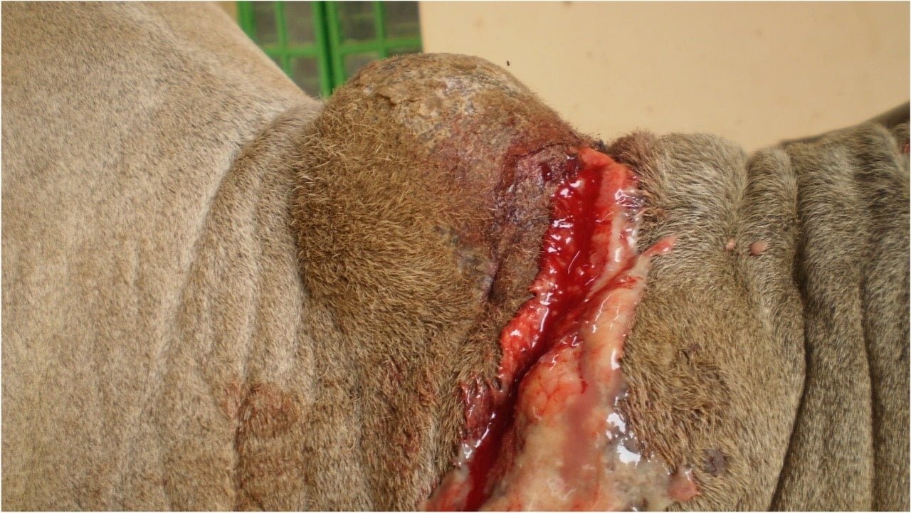
Yoke tumor may be a cystic swelling due to bursitis or a tumour mass due to chronic inflammation in the yoke region.
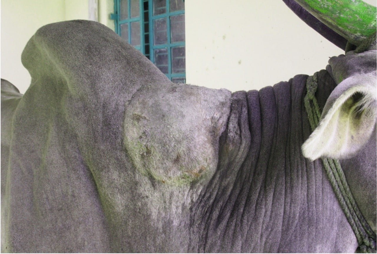
When the tumor is very large and involves most of the neck due to deposition of much fibrous tissue it is called tumour neck. If infection gains entry in to the swelling through superficial wound or injury this yoke gall converts in to abscess.
Yoke ulcer resulting from superficial abscess on the swelling.
Etiology
- Uneven and undue pressure of the yoke on the neck and its sudden movements are the causes
- Anything that weakens the neck on the yoke place.
- Thin neck (lean muscles)
- Young age (tender skin)
- Early castration (neck muscles fail to develop)
- General debility (decreases the resistance of neck muscles)
Favouring causes
- Anything that injure or contuses the yoke place
- Yoke with a rough surface
- Moist condition of the skin due to rain etc.
- Nervous temperament of the animal. Responsible for sudden, undue and unusual movements of the animal and the yoke
- Heavy loads, improper adjustment of weights cause unusual pressure.
- Unsuitable pair
Pathogenesis
An acute lesion and become chronic due to treatment with insufficient rest and
repeated work in the busy agricultural season or Chronic lesion from the outset due to slight and repeated injuries by the yoke.
Acute to chronic
Inflammatory exudates (due to the injury by the yoke) accumulate in the yoke place. Poor vascularity of the part slows its absorption. So the lesion requires treatment with longer rest to the part. But agricultural seasons forces work on the part before it becomes completely normal. This agains makes the lesion acute. Insufficient rest (during treatment) and work, alternately, for some time ultimately makes it chronic.
The exudates becomes organized, resulting in either local or diffuse fibrosis
This fibrosis effects the sensation of, and blood supply to the part. So it becomes unhealthy and easily excoriated.
Bacteria gain entire through the reaches into the area. Hot or cold, single or multiple abscess for
Due to constant irritation by the yoke, unhealthiness of the part, mobility and defective drainage results in ulcers.
Yoke Ulcer
Yoke ulcer is due to repeated injuries by the yoke. Much fibrous tissue is usually laid down. In long standing cases, the swelling reaches a large size and resembles a tumor – tumour neck.
Sometimes as a result of further contusion, the swelling becomes acute. Such acute phases may alternate periodically with phases of comparative quiescence during most of the animal’s working life.
Symptoms
- When acute those of acute inflammatory, gall, contusion cyst in yoke place.
- Occurrence of to swelling is sudden. It may be small or as large as a foot ball.
- Extension and flexion of neck is prevented in very severe cases.
When chronic- those of chronic inflammation, localized or diffuse fibrosis, unhealthy skin, small cold indurated abscesses in the subcutis, multiple sinnses and indolent ulcers in the yoke place.
Prognosis
Prognosis of yoke ulcer in early stages favourable. The exudates gets absorbed in one or two weeks.
Treatment
For acute lesion: Paint liquor iodine. Apply acetic acid chalk paste or kaolin paste or Magnesium Sulphate and glycerin paste for a few days, until the part becomes normal.
For cystic swellings, acute and chronic abscesses on general principles.
For multiple cold abscesses with unhealthy skin: Blistering the region and opening them after maturation and treating on general lines.
For solitary cold abscess with healthy skin: Enucleation with its walls intact, aseptically as in operative surgery guide. The incision should never be across or along, but oblique to the neck. It should not be on the mid dorsal line of the neck. Aim first intention healing. The animal must be put to work, 3 or 4 weeks after the removal of sutures, gradually to avoid the rupture of the embryonic tissue in the operation site.
Affections of Wither
Wither is the region over the backline where the neck joins the thorax and where the dorsal margins of the scapula lie just below the skin.
Fistulous withers (supraspinous bursitis) and poll evil are the surgical affections of Wither.
Fistulous withers and poll evil are rare, inflammatory conditions of horses that differ essentially only in their location in the respective supraspinous and supra-atlantal bursae.
Etiology
- Infection – mainly through the organism Brucella abortus found near cattle
- Streptococcus zooepidemicus, Actinomyces bovis, occasionally B suis
- Parasites (Onchocera cervicalis)
- Trauma to the area
- Fitting saddles
- Overwork
- Overloading
- Badly balanced loads
- The organism Brucella abortus, normally found in cattle, is the main cause of fistulous withers. The organism enters the horse’s body through an orifice i.e. the mouth, nose or eyes, or through broken skin.
Symptoms
- Swelling of the withers
- Heat in the withers
- Holes and tracts in the withers
- Build up of fluid at the withers
- Drainage in the form of a yellow/clear ooze
- Signs of fever and pain
- Sinus infection type symptoms
- Harness sores
- Hair loss
- In the early stage of the disease, a fistula is not present. When the bursal sac ruptures or when it is opened for surgical drainage and secondary infection with pyogenic bacteria occurs, it usually assumes a true fistulous character.
- The inflammation leads to considerable thickening of the bursa wall. The bursal sacs are distended and may rupture when the sac has little covering support. In more chronic, advanced cases, the ligament and the dorsal vertebral spines are affected, and occasionally these structures necrose.
- In the early stage, the supraspinous bursa distends with a clear, straw-colored, viscid exudate. The swelling may be dorsal, unilateral, or bilateral, depending on the arrangement of the bursal sacs between the tissue layers. It is an exudative process from the beginning, but no true suppuration or secondary infection occurs until the bursa ruptures or is opened
Diagnosis
- Clinical signs
- X ray – indicating presence of osteomyelitis of the underlying spinous process
- Ultrasound
Treatment
First remove the irritant and keeps the animal as quiet as possible, to prevent the muscles moving on each other.
If the skin is not broken and the swelling appears tense, hot and painful, cold applications may be applied to try and reduce the inflammation and the swelling. These may be in the form of cold-water irrigation and cooling lotions applied by soaking linen cloths and placing them across the wither.
If in the course of a few days the swelling does not disappear and the pain subsides, but on the contrary continue to increase, indicating suppuration. For this hot water fomentations must be diligently applied, together with some stimulating liniment, such as that of ammonia and turpentine.
It is an old but sometimes successful practice to “plug” the sinus to the very bottom with some caustic, such as corrosive sublimate, or arsenic, or a mixture of the two. This destroys the tissues for some distance around, and frequently brings away the damaged structure that prevented healing in the first instance.
Finally surgical correction should be carried out under general anaesthesia. An incision should be made at the lowest part of the cavity, so as to give free exit to the matter (pus) and allow of the removal of any dead tissues that may exist, and drainage of the abscess may be effected by passing a piece of tape (seton) through the wound, being careful to bring it out at a lower level than the floor of the cavity, so that no matter may be allowed to accumulate there. Care should be taken to avoid penetration of the dorsoscapulat ligament.
Sometimes the pus will have burrowed behind the shoulder-blade, in which case a depending opening must be made or a seton passed through it. At other times the projections of the backbones (vertebral spines) will be diseased, in which case they must be freely scraped or removed by the veterinary surgeon.
First day the cavity should be flushed with irritant antiseptic (eg. tincture iodine) and second day use non-irritant solution (eg. Povidone iodine).
If the dead space is more means the cavity should be packed with antiseptic gauze and it should be removed every alternative day. • Once it starts healing use antiseptic spray with fly repellent.
Prevention
Always have well-padded and properly-fitting harness and clothing, and as soon as any sign of chafing occurs, at once remove the offending agent.
In this way many tedious and painful wounds may be avoided.
It is reasonable to keep horses separate from Brucella -infected cattle, and cattle separate from horses with discharging fistulous withers.
Prognosis
It will cure if treated early. Suppuration causes arthritis of the intervertebral joints, extend to the spinal cord causing death.