TABLE OF CONTENTS
Surgical affections of lips
Surgical affections of lips in animals are foreign bodies, laceration, Avulsion, infections, eosinophilic ulcer disease, facial nerve paralysis, cleft lip, burns and neoplasms.
Anatomy of Normal lips
The lips of animals form the rostral and most of the lateral boundaries of the mouth and are separated from the upper and lower dental arcades by the vestibule.
The upper and lower lips form the oral fissure and meet at posterior angles, with the commissures. Except for the rostral two thirds of the upper lip, there is no hair along the lip margins.
Conical papillae are present on the caudal margin of the lower lip. The mucosa of the lower lip is firmly attached to the gum between the canine and first premolar teeth at the interdental spaces.
The philtrum is the deep, narrow cleft between the two halves of the upper lip.
In large animals lips play as a prehensile organ also.In bovines they are less mobile. In sheep the philtrum is more deep. In camel the upper lip is divided in to two indepedent halves by a deep fissure.
Muscles of lips are orbicular muscle for the voluntary movement, dorsal and ventral incisive muscles, maxillonasolabial, buccinator, zygomatic, and levator nasolabial for voluntary lip movement.
The musculature of the lips is innervated by the dorsal buccal, the ventral buccal, and the auriculopalpebral branches of the facial nerves.
Physiology of Normal lips
lips of animals contribute little to active prehension (In horses lips are the prehensile organ) and displays behaviour, including threatening attitudes. Scent marking through the application of secretions from glands.
Diseases or affections of lips
- Foreign Bodies
- Lacerations
- Avulsion
- Eosinophilic Ulcer Disease
- Facial nerve paralysis
- Harelip (Chiloschisis)
Foreign Bodies in lips
Foreign Bodies in lips of animals are Grass pieces, splinters of bone quills, wood pieces, bullets and carbon particles are usually seen. Sometimes pieces from dog chain and belts are also reported.
The animal attempts to expel or encapsulate the foreign material results in open wound followed by infection with bacteria. Further injury will be caused when the animal attempts to dislodge the foreign body.
Clinical signs of Foreign Body
Clinical signs of Foreign Bodies in lips of animals are-
- Inappetance
- Ptyalism (drooping of saliva)
- Dullness and pain
- Presence of an infected wound
- Direct visualization of foreign body under illumination
Treatment of Foreign Body
Foreign body can be removed with forceps. If the foreign body is buried deep within the tissue, general anaesthesia, routine surgical preparation of the site, and an incision in adjacent healthy skin or mucous membrane to remove the object.
The wound is then gently flushed with warm sterile saline, the skin or mucous membrane incision is closed. Small opening should be left open for drainage.
Post operatively antibiotics and anti-inflammatory drugs are indicated for 5 days. Oral antiseptic gel and soft light food till wound heals.
Lacerations of lips
Etiology of Lacerations
Etiology of Lacerations of lips in animals are Fights , broken edged objects and other injuries on lips.
Clinical Signs of Lacerations
Clinical Signs of Lacerations on lips are Ptyalism dullness and pain, Wound with irregular edges.
Treatment of Lacerations
Patient should be restrained before surgical repair. Haemostasis and debridement in addition to antibiotic therapy is usually initiated until the severity of the damage is assessed.
To close lip wounds, simple interrupted absorbable sutures are placed in the muscle and submucosa.
The mucosa is not included in the suture, and the knots lie deep in the lip tissue. Repair of large lip defects needs a skin flap or graft .The overlying skin is closed with simple interrupted sutures or vertical mattress sutures.
Post operative antibiotics and anti-inflammatory drugs for 5 days Soft light food till wound heals. Suture removal is recommended after 10-14 days.
Avulsions of lips

Etiology of Avulsions
Etiology of Avulsions of lips in animals are Automobile accidents and falls from heights.
Treatment of Avulsions
Suture the lip in place in minor avulsions with fine monofilament nylon or polypropylene. suture the interdental spaces or the teeth must be used to anchor the sutures if a large lip flap is displaced .
Mandibular symphysis should be examined and stabilised if separated with a full cerclage wire of 20 gauge.
Chelitis
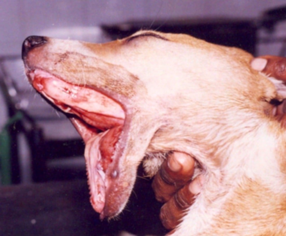
Chelitis is the inflammation of lips. this cause Lip fold pyoderma or intertriginous dermatitis.
Chelitis is common in spaniels, setters, and other breeds of dogs with large pendulous upper lips.

Pendulous lower lip in a great dane species of dog causing salivary drooling.
Etiology of Chelitis
Chelitis in animals caused by Bacterial, Viral, Canine viral oral papillomatosis, yeast and fungal candidiasis, dermatophytosis, coccidioidomycosis blastomycosis, cryptococcosis nocardiosis.
Treatment of Chelitis
Bacterial Dental prophylaxis: Daily cleansing of the lip folds with 2.5% benzoyl peroxide shampoo until the condition improves followed by maintenance cleansing every 2 to 5 days. Surgical extirpation of the folds to remove a lateral lip fold an elliptical skin incision encompassing all infected tissue and a margin of healthy tissue is made around the fold.
The dermis and subcutaneous tissues are undermined to remove all involved tissue.
The lateral lip fold is incised covering the infected tissue and a margin of healthy tissue. After removing the fold the edges of the wound are undermined to allow skin apposition to the mucocutaneous border without tension.
Papilloma Warts may be removed by sharp dissection at the level of their base with an electric scalpel. Spontaneous regression of the remaining warts usually occurs due to auto vaccination.
If Chelitis is fungal origin, Antifungal therapy should be given.
Eosinophilic Ulcer Disease
Eosinophilic Ulcer Disease is manifested as a well-circumscribed, red-brown, ulcerated, alopecic, glistening area on the skin of the lips or mucosa of the oral cavity.
Diagnosis of Eosinophilic Ulcer Disease in animals is based on history, physical examination, skin biopsy impression smear of the lesion.
Treatment is surgical excision or debridement of granulomas is difficult because of the paucity of surrounding tissue to use to repair defects. Deformity and recurrence are common complications.
Glucocorticoids and, in refractory cases, radiation therapy are the current recommended treatments.
Facial nerve paralysis
The facial nerve, supplies motor fibers to muscles of the face. Facial paralysis mostly affects motor function and except for taste, there is no loss of sensation from the skin and mucous membranes.
Chronic paralysis leads to facial muscle atrophy in animals.
Etiology of Facial nerve paralysis
Causes of Facial nerve paralysis secondary to direct nerve injury, space-occupying lesions, otitis media, and neuromuscular or central nervous system disease
Signs of facial paralysis are asymmetry of the ears, eyelids, and nose One ear may droop lip droops and saliva escapes from one corner of the mouth.
Nose and philtrum are drawn toward the unaffected side ocular fissure on the affected side is larger than normal and corneal and palpebral reflexes do not cause its closure.
Treatment of Facial nerve paralysis
Muscle nerve stimulation and Palliative surgery (to prevent drooling, a chelioplastic surgery) can be carried out.
Harelip (Chiloschisis)
Harelip or Chiloschisis is the cleft lip present in the upper lip. It may be unilateral or bilateral.
Cheiloplasty is the surgery performed for harelip in animals as reconstructive surgery under General Anaesthesia.
With the animal on its back the lower lip is pulled down to expose the lower incisor teeth . An incision is made along the mucogingival junction from the first premolar tooth on one side to the first premolar tooth on the other side.
The subcutaneous tissue is stripped from the mandible using a periosteal elevator.
The tightness of the lip determines the extent of dissection required. The lip should hang just ventral to the mucogingival junction.
If it doesn’t, additional length of lip should be dissected from the mandible. No sutures are placed.
The owner is advised to run their finger around the created pocket between the lip and mandible daily.
This has to be done to prevent the healing tissues from pulling the lip back into normal position. The wound heals by secondary intention healing.