TABLE OF CONTENTS
Surgical affections of guttural pouch
Surgical affections of guttural pouch in horses are guttural pouch tympany or Tympanites, empyema, mycosis, neoplasia and cyst.
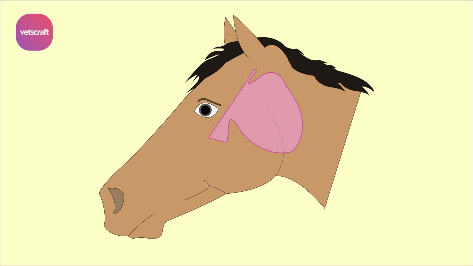
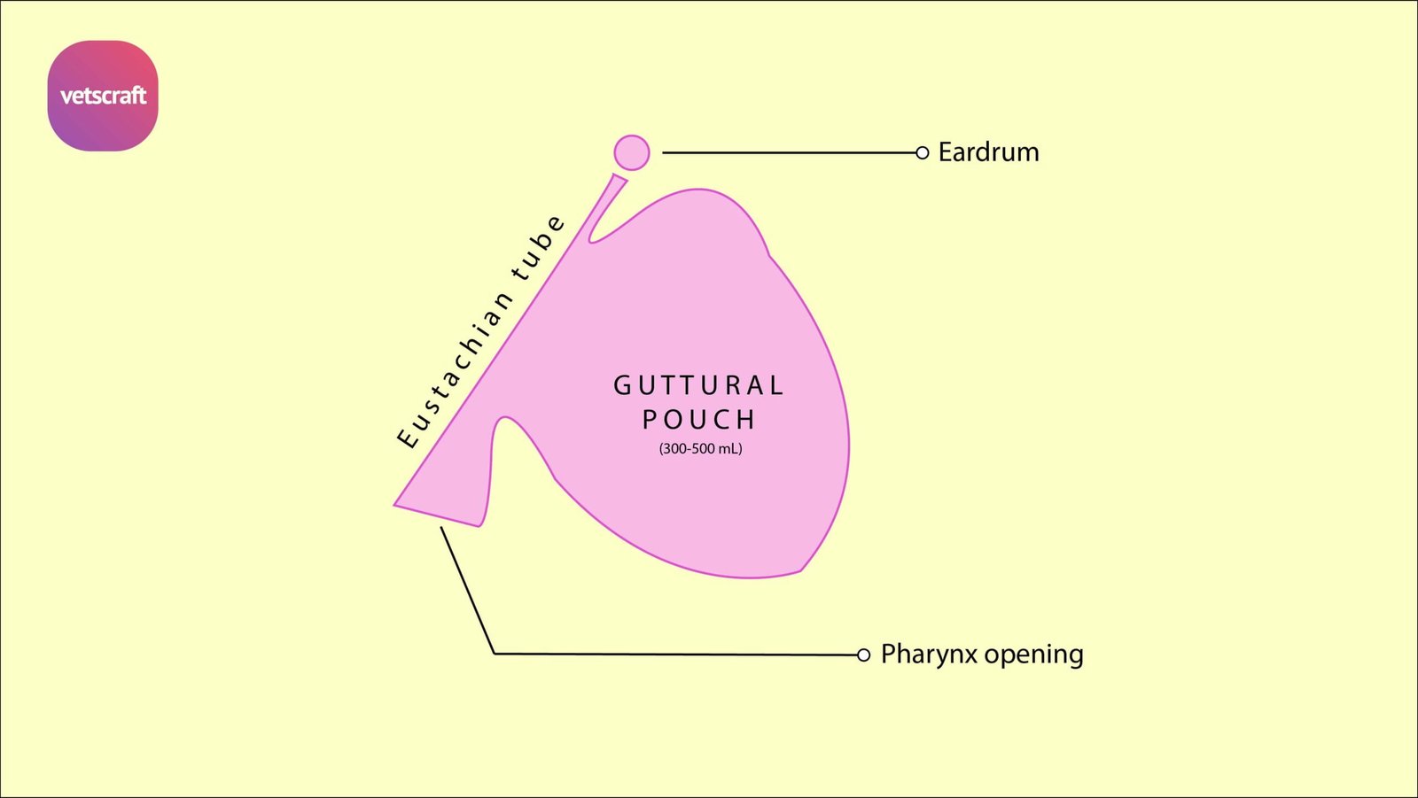
The guttural pouches are ventral invagination of the membranous part of auditory tube or Eustachian tube.

The capacity of each pouch approximates 300-500 mL. It is divided into a medial and lateral compartment by the invagination of the stylohyoid bone.
The mucosal lining of each pouch is secretory and is being covered by ciliated pseudo stratified epithelium with goblet cells and gland. The mucosal lining is generally thinner than that of nasopharynx.

Surgical affections of guttural pouch in horses
Surgical affections of guttural pouch in horses are-
- Guttural pouch tympany or tympanites
- Guttural pouch empyema
- Guttural pouch mycosis
- Guttural pouch neoplasia
- Guttural pouch cyst
Guttural pouch tympany or tympanites
Guttural pouch tympany or tympanites is a non-painful distention of the guttural pouch in horses with air characterized by an external swelling in the parotid region.
It is usually observed in young foals, although horses up to 20 months of age may be considered for this disorder. It appears to be more prevalent in fillies than in colts.
Usually only one guttural pouch is affected but it may occur bilaterally.
Etiology of guttural pouch tympany
The etiology of guttural pouch tympany of air accumulation within the pouch is not known but it seems that the air apparently enters the pouches during expiration or when the animal is swallowing.
In some cases infection is present in the affected pouch and the distension may be due to the formation of gas.
It has also been proposed that an abnormal mucosal flap at the pharyngeal orifice functions as a unidirectional valve trapping air or fluid within the pouch.
Clinical signs of guttural pouch tympany
Clinical signs of guttural pouch tympany depend on the degree of distension but the usual signs are a diffuse, painless, elastic tympanic swelling in the parotid region.
It may extend downwards and backwards towards the throat and upper part of the jugular furrow. If markedly affected, the foal may exhibit stertorous breathing, nasal discharge, Dysphagia, respiratory distress or evidence of pneumonia secondary to aspiration.
Pressure on the pouch may cause some of the air or gas to escape with a whistling sound.
Diagnosis of guttural pouch tympany
Diagnosis of guttural pouch tympany is based on significant clinical signs and radiographic examination of the skull and pharynx. X-ray reveals a large air filled guttural pouch with or without fluid accumulation.
Needle aspiration of air from one guttural pouch nearly correct the problem if a unilateral tympany exists.
Dorsoventral radiograph views may also be helpful in detecting bilateral involvement.
Treatment of guttural pouch tympany
Medical treatment like application of counterirritant or other topics to the skin over the pouch has no beneficial result. Puncturing the sac gives only temporary relief, as it rapidly refills.
Surgical correction is the good management of the guttural pouch tympany.
For unilateral tympany, surgical ablation of the median septum separating the two guttural pouches is performed. Bilateral involvement may necessitate resection of the excessive plica salpingopharyngeal flap.
Guttural pouch empyema
Guttural pouch empyema is the accumulation of exudates or pus within the guttural pouch and is usually a sequela of upper respiratory tract infection (Streptococcal species).
Etiology of guttural pouch empyema
Guttural pouch empyema may result from the rupture of abscessed retropharyngeal lymphnodes into the pouches or may accompany cases of guttural pouch tympany.
It may occur during the course of an infectious disease like influenza or strangles. Infection of the pouches usually becomes chronic because of lack of complete drainage.
It may occur usually secondary to an acute pharyngitis or may also accompany infectious parotiditis.
It may be associated with a neoplasm on its wall. Food material may enter the pouch through the Eustachian tube and give rise to suppurative inflammation.
Clinical signs of guttural pouch empyema
Signs include an intermittent white non-odorous nasal discharge (either unilateral or bilateral), lymphadenopathy, painful distention in the parotid region, stertorous breathing , Dysphagia, swelling of the submaxillary lymphatic glands, a rattling noise in the pouch during exercise, interference with swallowing and respiration, rupture of the pouch rare occurrence and occasionally epistaxis.
Inspissation of the material may occur with chronic infections, forming masses called Chondroids
Chondroids
If pus has been retained in the guttural pouch for a long time, much of the fluid portion drains and the inspissated portion remains as concretions commonly called as Chondroids.
Diagnosis of guttural pouch empyema
Diagnosis of guttural pouch empyema is based on clinical findings and confirmed by radiographic examination. Radiograph reveals a fluid line or opacity in the pouch. Inspissated material may also be evident radiographically.
On endoscopic examination, a purulent material may be seen at the pharyngeal orifice of the Eustachian tubes.
Prognosis of guttural pouch empyema
Prognosis is unfavourable i.e. there is no chance of spontaneous cure but when the pus becomes inspissated and the quantity of fluid is diminished the functional disturbance is relieved.
Death rarely supervenes from haemorrhage due to ulceration of the mucous membrane. Some cases prognosis is favourable when complete drainage and removal of contents is performed.
Treatment of guttural pouch empyema
Treatment of guttural pouch empyema may entail both medical and surgical modalities but opening and draining the guttural pouch is the only successful method of treatment for the accumulated pus.
Medical treatment includes lavage of the guttural pouch with saline solution and administration of systemic antibiotic is an initial step in therapeutic management.
Antiseptic inhalation, which may be of some use in a recent case by reducing inflammation of mucous membrane. The passage of Gunther’s catheter to evacuate the fluid contents and to enable the cavity to be irrigated with an antiseptic lotion introduced through the catheter by means of a syringe.
Surgery may be carried out if medical therapy is unsuccessful or if the material within the guttural pouch is inspissated.
Surgical procedure for guttural pouch empyema (Hyovertebrotomy Viborg’s triangle)
Site of operation: Antero-inferior border of the wing of the atlas.
Make an incision about three inches long antero-inferior border of the wing of the atlas, going through the skin without making dissection the parotid gland. Reflect the gland forward by blunt dissection of the loose connective tissue beneath it.
Separate the areolar tissue, digastricas, stylo-maxillary and occipito-styloid muscle until the pale lining of the pouch is visible. Grasp a fold of the guttural pouch membrane with an artery forceps and incise it.
Enlarge the opening thus made with the fingers or the jaws of a forceps and the interior of the pouch will then be quite visible. Evacuate the contents which may be entirely liquid or partly solid in the form of chestnut-like bodies called chondroids.
To provide better drainage, a counter opening is made in the center of Viborg’s triangle. This is defined as the tendon of sternomaxillaris muscle, the sub maxillary vein and the caudal border of the vertical ramus of the mandible.
Pass a stout metal sound into the pouch and make it bulge the skin in the center of the triangle. Keep this opening patent for a few days by inserting a strip of gauze through it and the upper opening.
The surgical wound heals by granulation.
Guttural pouch mycosis
Guttural pouch mycosis is caused by Aspergillus. Multiple or diffuse fungal plaques on the caudodorsal aspect of the medial guttural pouch is the common site of lesion.
Clinical signs of guttural pouch mycosis
- Unilateral or bilateral epistaxis
- Epistaxis
- Abnormal head extension
- Ocular changes
- Facial nerve paralysis
- Lingual hemiplegia
Treatment of guttural pouch mycosis
Treatment of guttural pouch mycosis is anti-fungal agents and surgical interventions if necessary.
Guttural pouch neoplasia
Guttural pouch tumors are rare. The most common is melanoma, usually seen in gray horses. Tumors in the guttural pouch may cause neck swelling and interfere with the nerves and vessels that run through the pouch. This may result in epistaxis, difficulty swallowing etc.
Guttural pouch cyst
Guttural pouch cyst in horses are very rare. these are fluid containing capsules in the guttural pouch.