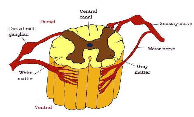
- The spinal cord is that part of the central nervous system situated in the vertebral canal. It extends from the foramen magnum to about the middle of the sacrum. It is approximately cylindrical and more or less compressed from above downward. It is continuous with the medulla oblongata at the foramen magnum, where there is no natural line of demarcation. Its terminal part rapidly narrows to a point called the conus medullaris. This is prolonged for a short distance by a slender filament of piamater called filum terminale.
- As a result of the unequal growth of the nervous tissue and the vertebral column, correspondence between the two is not exact and the spinal nerves usually have to travel a short distance backward before they gain exit from the canal. The conus medullaris reaches only the anterior part of the sacral canal and hence the rest of the sacral canal is occupied by the sacral and coccygeal nerves extending backward in the canal for a considerable distance before they pass out of it, forming a sort of a bundle in the middle of which is the conus medullaris and filum terminale. This arrangement is termed as the cauda equina.

- The spinal cord is fairly uniform in the greater part of the thoracic region but there are two conspicuous enlargements that involve segments with which the nerves of the limbs are connected. The cervical enlargement begins about the fifth cervical segment and subsides at the second thoracic and the lumbar enlargement is at about the fifth lumbar, beyond which the cord rapidly narrows to form the conus medullaris.
- The surface of the cord is divided into two equal halves by a dorsal median groove and a ventral median fissure. On either side of the former is a dorso-lateral groove where the dorsal root fibres of spinal nerves enter the cord. The ventral root fibres leave the cord a little lateral to the ventral median fissure. The dorsal median groove is filled up by the dorsal median septum and the ventral one is narrow and deep and reaches to about the middle of the cord. The two halves of the cord are connected by commissures of gray and white mater. The gray commissure is a transverse band at the base of the dorsal median septum and is traversed by the central canal of the spinal cord. The white commissure is a bridge of white mater that connects the two ventral columns of white mater.
- The central canal opens anteriorly into the posterior angle of the fourth ventricle behind it ends in the conus medullaris forming a slight dilatation, the ventricularis terminalis. It is lined by ciliated epithelium.

TABLE OF CONTENTS
GRAY MATER
- The gray mater of the spinal cord in transverse section resembles roughly the capital letter ‘H.’
- The cross bar being the gray commissure. Each half consists of a dorsal and a ventral horn.
- The dorsal horn is elongated, narrow and reaches the surface of the cord at the dorsolateral fissure at the entrance of the dorsal root fibres. It contains sensory neurons.
- The apex of the dorsal column is covered by a mass of translucent nervous tissue called substantia gelatinosa that contains cell bodies in the pain and thermal pathways. From the middle of the cervical region to the lumbar region, there is a medial projection of gray mater at the ventral part of the dorsal column known as nucleus dorsalis.
- A pointed projection from the lateral surface of gray mater, opposite the gray commissure is known as lateral horn or intermedio-lateral column and is seen only in the thoracic, lumbar and middle sacral levels. It contains autonomic motor neurons.
- The ventral horn is rounded, wider and is separated from the surface of the cord by the white mater through which pass the ventral root fibres. It contains somatic motor neurons.
WHITE MATER
- The white mater is divided into three pairs of columns.
- The dorsal column lies on either side of the dorsal median septum to the dorsolateral groove.
- The ventral column is situated between the ventral median fissure and the ventral root. These are connected by the white commissure.
- The lateral columns are included between the dorsal and ventral root fibres. The amount of gray and white mater differs in different regions.
- The gray mater increases from the cervical to the sacral region.
- The white mater consists chiefly of medullated nerve fibres arranged in three columns or funiculi. Some of the fibres cross in the median plane to the opposite side through the white commissure but the majority directed to the same side.
- The latter are chiefly of three classes
- Those which carry impulses from the periphery to the centre called ascending sensory or afferent tracts
- Those which carry impulses from the centre to the periphery – descending motor or efferent tracts
- Those which connect the different segments of the cord – the intersegmental tract.
- The nerve tracts or fasciculi are not recognizable in the natural state but their identity has been established by special methods.
- The spinal cord of a medium sized ox is about 1.6 to 1.7 m long and weighs 2-2.5kg.