TABLE OF CONTENTS
Sternum Bone of Domestic Animals
The sternum of domestic animals is a medially placed, segmented bone that forms the ventral (floor) boundary of the thoracic cavity.
Sternum bone play role in protecting the underlying thoracic organs, such as the heart and lungs, and provides articulation points for the costal cartilages of the ribs.
Sternum Bone of Ox
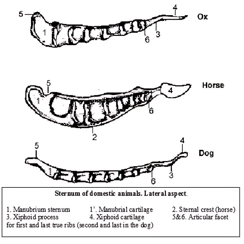
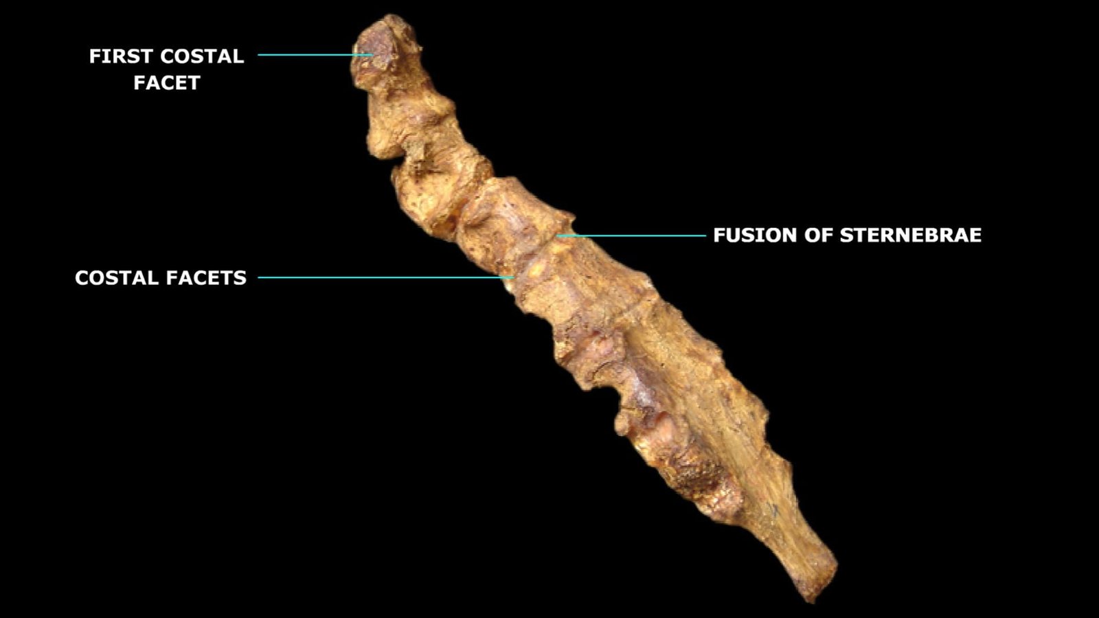
The sternum of the ox is a medially placed, segmented bone that forms the floor of the thorax. It is made up of seven bony segments, called sternebrae, and is elongated from front to back.
The segments are connected by intervening cartilages. It is compressed laterally until about its middle, and thereafter it is flattened from above downwards. It is directed obliquely downwards and backwards.

The sternum of the ox presents two surfaces, two borders, and two extremities.
The dorsal face is narrow in front but gradually becomes wider towards the back. This face provides attachment for the internal ligament in the middle and the transversus thoracis muscle laterally. It forms the floor of the thorax.
The ventral face is extensive and is strongly convex in front but more or less concave behind the second sternebra. This surface provides attachments for the pectoral muscles and the rectus abdominis muscles. The two surfaces are marked by transverse lines indicating the lines of fusion of the sternebrae.
The lateral borders are thick in front but become thinner gradually towards the back.
Each border presents eight articular cavities for articulation with the ventral extremities of the first eight costal cartilages. The first of these is on the anterior part of the dorso-lateral aspect of the first sternebra. Except for the first and the last two, the other cavities are located between the sternebrae. The facets for the seventh and eighth costal cartilages often become confluent.
The anterior extremity is the anterior end of the first sternebra, which forms the manubrium sterni or presternum. The posterior end of this segment articulates with the anterior end of the second sternebra to form a diarthrodial joint. The anterior extremity provides attachment to the sternocephalicus and sternothyrohyoideus muscles.
The posterior extremity, or metasternum, is formed by the xiphoid cartilage, which is thin, flexible, and nearly circular. Between the pre- and metasternum lies the body or mesosternum.
The diaphragm is attached across the upper concave face of the xiphoid cartilage, close to its junction with the last sternebra. The part of the cartilage behind the line of attachment of the diaphragm forms the floor of the abdomen. The ventral face provides attachment to the transverse abdominis and linea alba.
Comparative Anatomy of Sternum Bone
The sternum varies among domestic animals in shape, size, and number of segments. While most mammals like ox, sheep, and pig have a segmented sternum with six to eight sternebrae, birds like fowl have a large, keeled sternum to support flight muscles.
Sheep and Goats
- The sternum of sheep and goats resembles that of the ox.
- The number of segments may be reduced to six.
- The first segment is cylindrical with enlarged ends.
- The second and third segments are wide and flat, while the last segment is long and narrow.
Horse
- The sternum of the horse is composed of seven sternebrae. It is canoe- or boat-shaped and presents three surfaces, three borders, and two extremities.
- The superior surface is narrow and triangular.
- The two lateral surfaces are convex above, slightly concave below, and present costal cavities on their upper parts.
- The dorsolateral borders separate the dorsal and lateral surfaces.
- The inferior border forms the keel.
- The anterior extremity presents a laterally compressed cariniform cartilage, which has a deep notch superiorly for articulation with the two first costal cartilages.
- The first sternebra is fused with the second, and the manubrium is made up of the cariniform cartilage and the first sternebra.
Pig
- The sternum of the pig consists of six segments and resembles that of the ox in general.
- The first segment is long and bears a blunt, pointed cartilage at its anterior end. Its posterior end forms a diarthrodial joint with the body.
- The last segment has a long, narrow part that bears the xiphoid cartilage.
Dog
- The sternum of the dog is composed of eight sternebrae, all of which are fused.
- Each sternebra is slightly compressed from the sides and constricted in the middle.
- The anterior end bears a small presternal cartilage, and the xiphoid cartilage is narrow.
Rabbit
- There are 6 sternebrae in the sternum of the rabbit.
- The body of the sternum extends from the 2nd to the 5th sternebra.
- The xiphisternum is the last sternebra, formed from the bony xiphoid process and the terminal spatula—the xiphoid cartilage.
Fowl
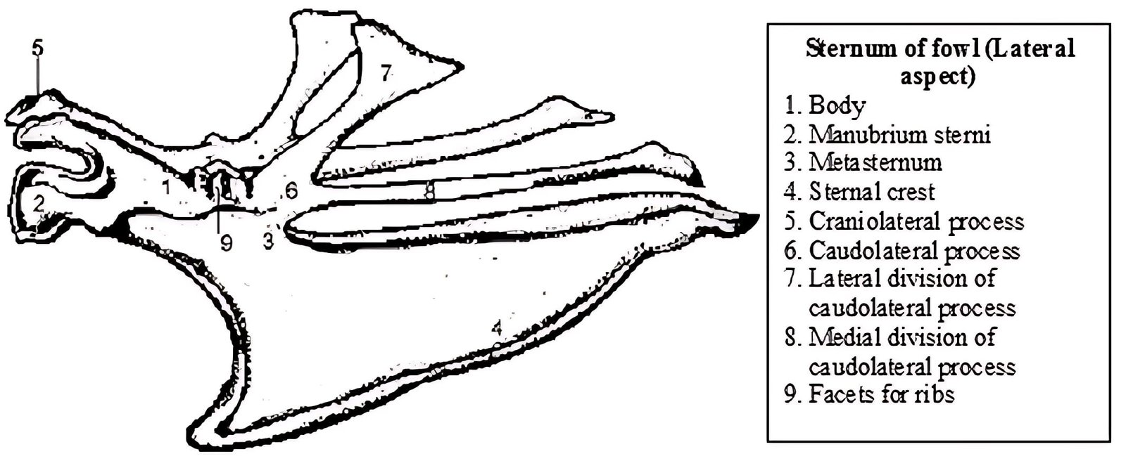
- The sternum of the fowl differs from the corresponding structure of domestic mammals since it is formed entirely of bone in the adult. It results from the fusion of five pieces, which have separate centers of ossification in the chick.
- The central wide part forms the body of the bone. In front of this is the anterior process—the rostrum or manubrium—and behind is the posterior process or metasternum.
- The dorsal surface of the body is concave and pierced by foramina, which permit air to enter the interior from the adjacent air sacs.
- The ventral surface is encroached upon by the lower borders of the anterior and posterior processes.
- The anterior process, or rostrum, is short, and on either side of its root is an elongated facet for articulation with the coracoid bone.
- The posterior process, or metasternum, is very long, being in fact more extensive than the body itself. It carries ventrally a thin plate of bone called the keel or sternal crest, which affords attachment to the pectoral muscles.
- The keel is broad in front and fades out posteriorly. United to each lateral border of the body are the anterolateral (costal) process in front and the posterolateral process behind. The costal process projects upwards and forwards.
- The posterolateral process is short and divides into medial and lateral xiphoid processes. The lateral division is broad, plate-like, and covers the last two or three ribs. The medial division is longer and projects backward, parallel to the lateral border of the metasternum. The deep notches between them and between the posterior division and metasternum are closed in life by a tough membrane.
- Between the anterolateral and posterolateral processes, the lateral border of the body shows four depressions for articulation with the third, fourth, fifth, and sixth ribs. Occasionally, the last rib also meets the sternum.