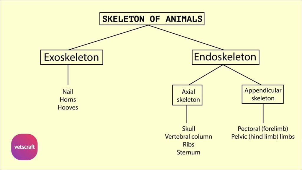
TABLE OF CONTENTS
Splanchnology of Thoracic cavity
The thoracic cavity is the second in size of the three body cavities. In form, it is somewhat like a truncated cone with the base cut off very obliquely and directed backwards
- Thoracic cavity presents a roof, floor, two lateral walls, a caudal wall and an cranial aperture or inlet
- The bodies of the thoracic vertebrae form the roof. The lateral walls are formed by the ribs and intercostal muscles. The sternum forms the floor
- The caudal wall is formed by the convex face of the diaphragm
- The inlet is small, narrow and oval. It is bounded above by first dorsal vertebra below by the first segment of the sternum and laterally by the two first ribs
- It is closed in life by structures passing into and out of the thorax viz., the longus colli muscles, the trachea, the oesophagus, vessels, nerves and lymph glands
- A longitudinal septum termed the mediastinum thoracis or septum mediastinale, extends from the dorsal wall to the ventral and caudal walls, and divides the cavity into two lateral chambers termed the pleural cavity. Each of these chambers is lined by the serous membrane, called the pleura
- The mediastinum is, for the most part, not median is position, as might be inferred from its name this is correlated with the fact that the largest organ contained in it the heart, is placed more on the left side than on the right consequently the right pleural cavity and lungs are larger than the left
- Practically all the thoracic organs are in the mediastinal space between the pleurae, with the exception of the lungs, caudal vena cava and part of the right phrenic nerve
- The part in which the heart and the pericardium are situated together with that dorsal portion is usually called the middle mediastinal space; the parts before and behind this are termed respectively the cranial and caudal mediastinal spaces
Species difference
Sheep and Goat
- It resembles that of ox
- The pleural sacs form a cul-de-sac on each side of the first lumbar vertebra
- Perforations are present in the cranial mediastinum of the sheep and it is not perforated in goat
Horse
- The thoracic cavity is larger
- The pleura are thin and the caudal mediastinum appears fenestrated and when these apertures are present the two-pleural sacs communicate with each other
- The apertures do not exist in the fetus
Pig
- The thorax is rounded due to strongly curved ribs
- The pleural sacs extend forward to the first intercostals space
- The diaphragmatic line of pleural reflection begins at or a little above the sternal end of the seventh and extends in a gentle curve to about the middle of the last rib
- When a fifteenth rib present, it does not affect the arrangement of the pleura of the diaphragm
Dog
- Thorax is proportionately larger
- Mediastinum is imperforate, but permeable to gas and water
- Both right and left cupola pleurae extend beyond the cranial border of the first rib
Rabbit
- General plan resembles the ox
Fowl
- Since the diaphragm is absent, there is no separate thoracic cavity
Pleurae
The pleurae are serous membranes, which enclose on each side a pleural cavity. These line the walls of the thorax from the lateral laminae of the mediastinum, and then reflected from the latter upon the surface of the lungs. Thus we distinguish parietal, mediastinal and pulmonary or visceral parts of pleura
- The parietal pleura line the thoracic walls. On the lateral thoracic wall it is adherent to ribs and intercostal muscles and is termed the costal pleura
- Behind it is closely attached to diaphragm, forming the diaphragmatic pleura
- The mediastinal pleura cover the organs in the mediastinal space and are in part in opposition with the opposite sac
- The part which is adherent to the pericardium is the pericardiac pleura. From the mediastinum, each pleura is reflected on the corresponding lung forming the pleura pulmonalis or the visceral pleura. The reflection occurs around and behind the hilus of the lung
- Caudal to the root of the each lung, there is a horizontal fold of pleura called the pulmonary segment
- The right pleura forms a special sagittal fold, which encloses the caudal vena cava in its upper edge; it is therefore called the fold of the vena cava (Plica venacava)
- It gives off a small accessory fold for the right phrenic nerve
- The pleural cavity contains a film of clear serous fluid, the liquor pleurae
- The cavity is normally a capillary space
- The pleura resemble the peritoneum in structure and appearance
- It is attached to the structures that it covers by a subserous tissue called endothoracic fascia
- The cupola pleurae or apex of each pleural sac lies at the thoracic inlet
- The right cupola projects beyond the cranial border of the first rib under the scalenus. The left cupola usually does not extend beyond the plane of first rib