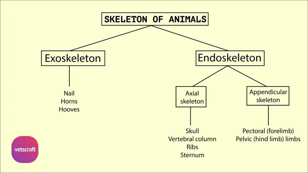
TABLE OF CONTENTS
Sphenoid Bone in Domestic Animals (Ox, Horse, Dog, Fowl) – Anatomy Explained
The sphenoid bone is a complex, unpaired bone located at the base of the skull in domestic animals. It plays a vital role in forming the floor of the cranial cavity and provides passage for important cranial nerves and blood vessels.
Structurally, it consists of two parts—the post-sphenoid and pre-sphenoid—each contributing to various foramina and sinuses. While its general form is conserved across species like ox, horse, dog, and fowl, notable anatomical variations exist that reflect differences in skull morphology and function.
Sphenoid Bone of Ox
The sphenoid bone of the ox is located at the base of the skull. In the calf at birth and for some months after, it consists of two pieces.
The posterior part, which lies next to the basilar part of the occipital bone, is termed the post-sphenoid, and the anterior part is the pre-sphenoid. Although the two parts fuse later in life, it is convenient to describe them separately.
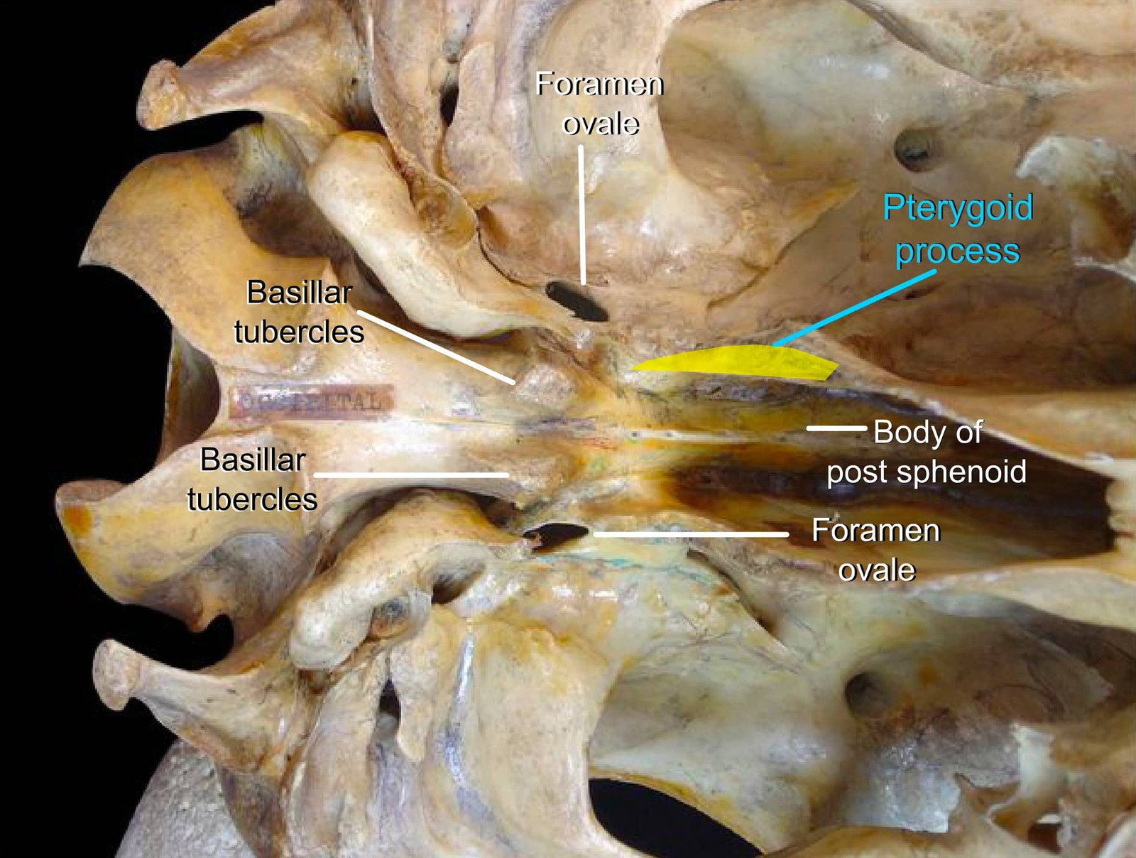
Post-sphenoid
The post-sphenoid has a body, two temporal wings, and two pterygoid (subsphenoidal) processes.
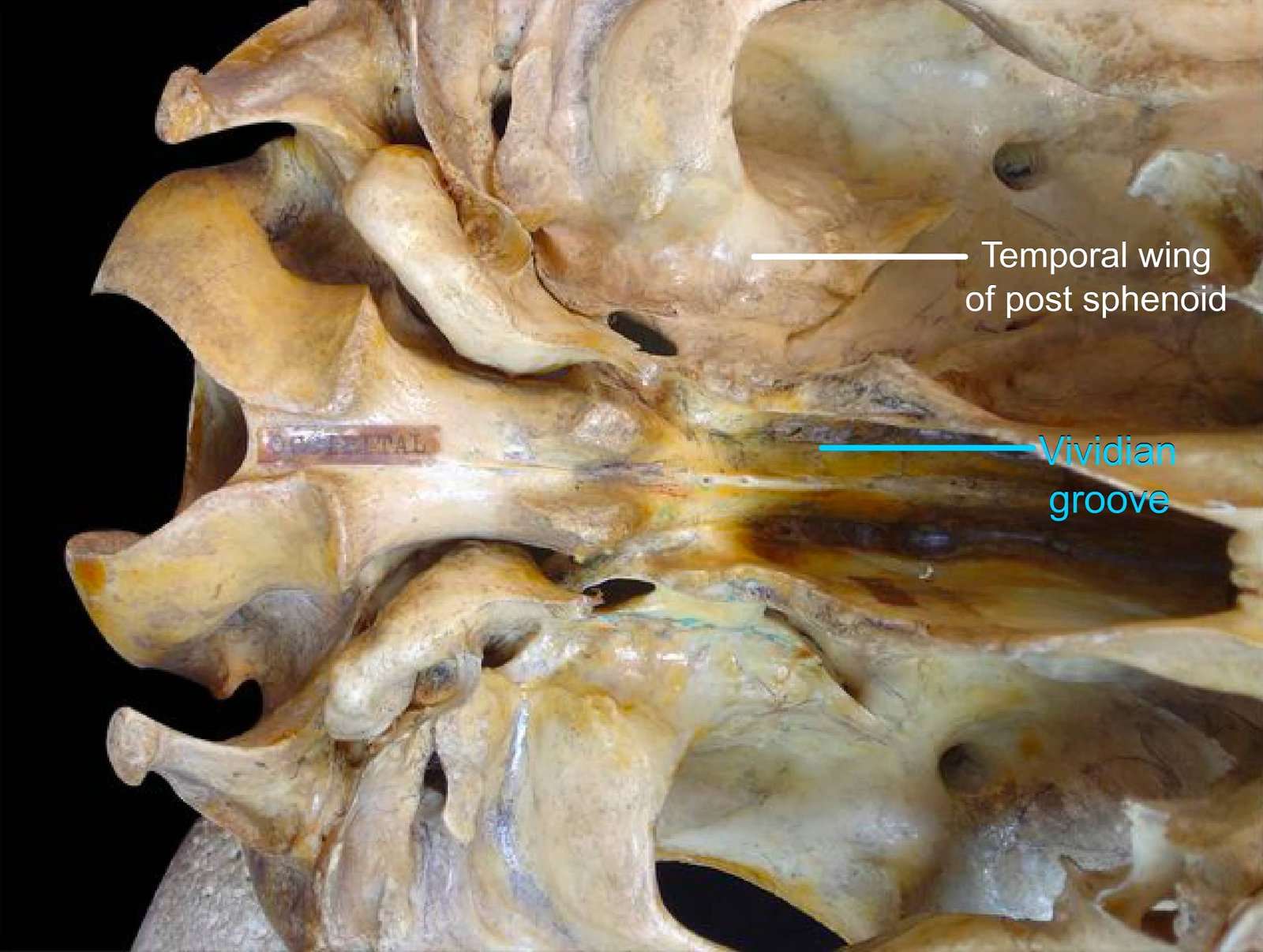
The external face of the body presents, close to its junction with the pterygoid, the vidian groove for the nerve of the pterygoid canal (vidian nerve).
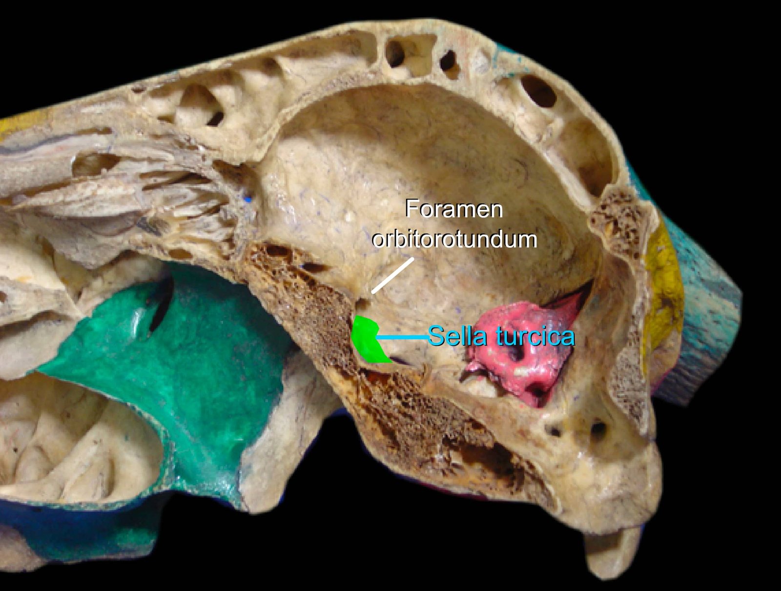
The internal surface presents the hypophyseal or pituitary fossa (sella turcica) for the pituitary gland. The dorsum sellae is a transverse projection at the posterior end of the body and bears posterior clinoid processes.
The wings diverge outward from the body. Each wing is perforated about its middle by the foramen ovale for the mandibular nerve and middle meningeal artery.
The internal surface of the wing presents a longitudinal groove leading to the foramen orbitorotundum. The posterior border forms the anterior margin of the foramen lacerum.
The anterior border presents a medial notch, which, together with a similar notch on the wing of the pre-sphenoid, forms the foramen orbitorotundum for cranial nerves III, IV, VI, and the maxillary and ophthalmic divisions of cranial nerve V.
It is free anterolaterally to form the pterygoid crest. The subsphenoidal or pterygoid processes spring from the anterior part of the wing and are directed downward and forward. Its medial face is covered by the pterygoid and palatine bones.
Pre-sphenoid
The pre-sphenoid lies at a higher level and has a body and two orbital wings. The anterior part of the external face of the body is mostly concealed by the vomer and laterally by the pterygoid bones.
The vidian groove is continued by a vidian canal at the junction of the wing with the body and opens into the pterygopalatine fossa.
The cranial surface of the body presents anteriorly a median ethmoid spine, which joins the crista galli of the ethmoid. Posteriorly, and at a lower level, is the optic groove, which supports the optic commissure in life, and the groove on either side leads to the optic foramen.
In the adult, the body of the pre-sphenoid is slightly excavated to form the sphenoidal sinus.
The orbital wings are larger than the temporal wings. They curve dorsolaterally from the sides of the body.
The external surface forms part of the orbital wall. It is overlapped by the frontal bone in such a manner that it appears to divide into two branches.
The anterior one of these joins the ethmoid and the perpendicular part of the palatine at the spheno-palatine foramen. At its junction with the body, it is pierced by the optic foramen.
The posterior border forms, with the wings of the post-sphenoid, the foramen orbito-rotundum. The internal face lodges the cerebrum.
Comparative Anatomy of Sphenoid Bone
The comparative anatomy of the sphenoid bone in domestic animals reveals key structural variations that are significant for species-specific functions.
Horse
- The posterior border of the wing of the post-sphenoid forms the anterior border of the foramen lacerum and presents three notches — carotid, oval, and spinous — from within outward.
- The foramen lacerum is a large triangular gap in the dry skull and is largely closed in life by dense fibrous tissue, with a posterior opening, i.e., the foramen lacerum posterius or jugular foramen, for the IX, X, and XI cranial nerves; and anteriorly, the notches referred to above are converted into the carotid artery, mandibular nerve, and middle meningeal artery, respectively.
- The dorsum sellae and post-clinoid processes are absent.
- The anterior border of the wing meets the wings of the pre-sphenoid. Above and below it, the free part forms the pterygoid crest, which continues on the pterygoid process.
- On or under the upper part of the crest, there is usually a small trochlear parvum, which communicates with the alar canal.
- The wing presents, close to its anterior border, the foramen rotundum (for the maxillary division of the V cranial nerve), and with the pre-sphenoid wing, it forms the foramen orbitale, which is separated from the foramen rotundum by a thin plate of bone.
- The foramen orbitale serves for the passage of the III, VI cranial, and ophthalmic division of the V cranial nerves.
- The pterygoid processes project downward and forward and curve outward at their lower part. The root of the process is perforated by the alar (subsphenoidal) canal for the passage of the internal maxillary artery.
- The wings of the pre-sphenoid form the foramen orbitale with the wings of the post-sphenoid. In front and above this is the optic foramen. Anteriorly, the wings meet the cribriform plate of the ethmoid and orbital plates of the frontal bone to form the ethmoidal foramen. The sphenoidal sinus in the body of the pre-sphenoid communicates with the palatine sinus in the vertical part of the palatine bone.
Dog
- The body of both the pre- and post-sphenoid is dorsoventrally flattened. The dorsum sellae and posterior clinoid processes are better developed in dogs.
- Anterior clinoid processes project from the roots of the orbital wings.
- The temporal wings are extensive and articulate with the parietals dorsally.
- The foramen orbitale is at the junction of the wings, a little lower than the optic foramen.
- An alar canal is present, and the foramen rotundum opens into it.
- The foramen ovale is as in the ox.
- The carotid notch, with a similar notch on the temporal bone, forms the carotid foramen.
- The sphenoidal sinus is absent.
Fowl
- The sphenoid is largely concealed by a large triangular basitemporal or sub-sphenoid bone in fowl.