Selenium and Vitamin E Deficiency
Selenium (Se) is necessary to protect cells against damage by lipid per oxidation and free radicals which are produced during normal cellular metabolism.
Selenium is a biochemical compound of the GSH-PX (Glutathione peroxidase enzyme). GSH-PX enzyme level in erythrocyte is directly relation to the blood concentration of the Selenium. This will aid in the diagnosis of the Selenium deficiency and determine the level of selenium in the tissue of the animal.
Selenium is also a component of thyroid gland to conversion of T4 to active T3 by iodothyroniniodinase (i.e. selenoprotein)
Vitamin E prevents hydrogen peroxide formation during lipid peroxidation. Vitamin E is combined with Selenium to maintain the integrity of the cell membrane, because Selenium has the sparing effect on Vitamin E.
Etiology
- Diet: Deficiency of Selenium or Vitamin E and or both
- High PUFA in diet: mostly it cause Vitamin E deficiency, because PUFA is the conditioning factor for Vitamin E.
Pathogenesis
- Selenium, Vit E and sulfur containing amino acid synergistically to protect tissues from oxidative damage.
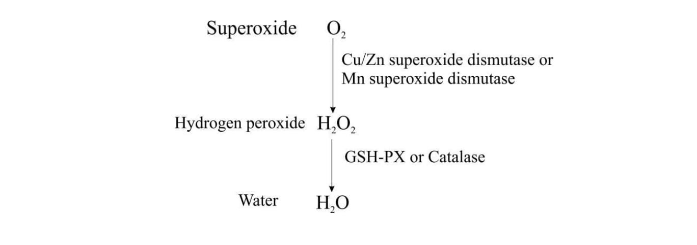
- GSH-PX is Selenium dependent, which is detoxifying the lipid peroxides and reduce to non-toxic hydroxyl fatty acids. GSH-PX inhibits the breakdown of H2O2 and certain organic peroxide product by glutathione during redox cycle. H2O2 and lipid peroxidation are capable of causing irreversible denaturation of essential cellular protein, which leads to degeneration and necrosis.
- Vitamin E prevents fatty acid H2O2 formation. High level of PUFA in the diet increases the requirement for Vit E.
- Low Vit E and Selenium in the diet leads tissue oxidation, which results degeneration and necrosis of the cells and tissues, especially liver, heart, skeletal muscle and blood vessels.
A balance of superoxide production with detoxification appears to be important to cellular health and intracellular metabolic signals. Superoxide to water conversion is as follows:
Selenium and Vitamin E deficiency causes different disease condition in various species as follows:
- Nutritional (enzootic) muscular dystrophy – lambs, calf, kid, piglets, horse
- Mulberry heart diseases – Pigs
- Hepatosis dieteticia – Pigs
- Exudative diathesis – Pigs
- Iron hypersensitivity – Pigs
- Ill thrift – Sheep
- Reproductive insufficiency – RFM – Cattle, Sheep
- Anemia – all species
Clinical Signs
Nutritional (enzootic) muscular dystrophy (NMD)
- Myocardial and diaphragmatic form:
- Young calves, lambs, foals
- It cause acute heart failure, respiratory distress and sudden death
- Skeletal form:
- Older animals – old calves, yearling cattle, older foals
- It cause weakness, recumbency and unresponsive to treatment
- Congenital form:
- Either sill birth or birth of weak calf, that fails to thrive and usually die within few days of birth.
- Acute enzootic NMD:
- Affected animal collapse and die suddenly after exercise without any signs
- Increased heart rate (up to 150-200/min) and respiratory rate (up to 60-72/min).
- Loud breath sound is audible over the lung field.
- Sub acute enzootic NMD:
- It most notice in rapidly growing calve – White muscle disease.
- Lambs – stiff lamb disease
Affected animal found in sternal recumbency and unable to stand, if stands with support animal shows stiffness, trembling of limbs and weakness
Laboured and abnormal respiration – due to involvement of diaphragmatic and intercostals muscles. Temperature is normal and sometimes transient fever (41 degree C) due to myoglobinuria.
Mulberry Heart disease
- Mostly in pigs – up to 4 weeks of age
- Found dead without any sign
- Severe dyspnea, cyanosis, recumbency and when animal make to walk t forcefully animal may die immediately.
Hepatic dietetic
- Mostly in pigs
- Found dead
- Dyspnea, depression, vomiting, staggering gait, diarrhea and collapse
- Icteric and pale mucus membrane
Reproductive defect
- RFM
- Infertility
Diagnosis
- Based on history
- Clinical signs
- Laboratory findings
Laboratory findings
- Plasma Creatinine kinase (CK)– Increased in NMD
- Increased AST
- Increases in both CK and AST is directly related to severe muscle damage.
- Selenium status in soli and plant
- Selenium status assessed by serum, whole blood and GSH-PX
- Blood and milk level of Selenium are also used to detect selenium deficiency
- GSH-PX level is directly relationship between the blood and tissue. Hence the estimation of GSH-PX is ideal for the confirmative diagnosis.
- Vitamin E status- alpha tocopherol level in blood and liver provide good information on Vitamin E. But estimation of Vit E is cost effective and not done commercially.
Treatment
Combination of Selenium and Vitamin E is used for the treatment, because of the overlapping function of the Selenium and Vitamin E.
Combination of 3 mg of sodium of potassium selenit and 150 IU/ml of DL- alpha tocopherol acetate in IM at 2ml/45kg body weight single administration.
Prevention and Control
- Provide dietary supplement with Selenium and Vitamin E (Selenium at 0.1mg /kg DM)
- Supplement during pregnancy
- Parenteral Selenium injection:
- Sodium selenite
- Selenium & Vitamin E injection
- Cattle & sheep: 1 mg of Se/kg body weight (3 weeks before breeding)
- Pigs: 0.05 0.1 mg of Se/kg body weight
- Oral Selenium and anthelmintic.
- Top dressing – deficient soil – Sodium selenit @ 10g/ha is effective for 12 month.
- Vitamin E @ 1000-3000 IU/ day

