TABLE OF CONTENTS
Salivary glands of different animals
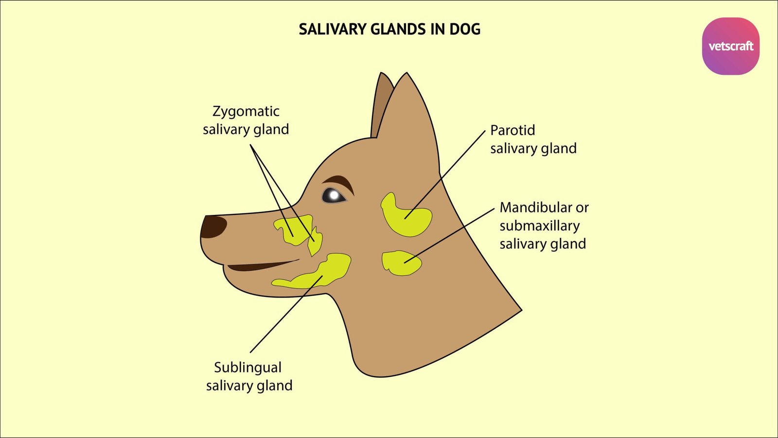
Salivary glands of different animals are Parotid, Mandibular or submaxillary, Sublingual and zygomatic salivary gland.
There are four pairs of salivary glands situated on the sides of the face and the adjacent part of the neck-
- Parotid salivary gland
- Mandibular or submaxillary salivary gland
- Sublingual salivary gland
- Zygomatic salivary gland

Of these salivary glands, the parotid salivary gland is of serous type, while the mandibular, sublingual and zygomatic salivary glands are of the mixed type.
Parotid salivary gland
Parotid salivary gland of Ox
The Parotid salivary gland of Ox is situated below the ear in the space between the ramus of the mandible and the wing of atlas, overlapping the caudal part of the masseter muscle.
Parotid salivary gland has the form of a very narrow triangle with its base upwards. It is light red brown in colour and weighs about 115 gm. It presents for description two surfaces, two borders, a base and an apex. The lateral surface is covered by the parotid fascia and the parotido-auricularis muscle.
Parotid salivary gland of Ox is crossed obliquely by the external jugular vein at its lower part. The medial surface is uneven and has many important relations -the great cornu of the hyoid bone, masseter, digastric and occipito-hyoideus muscles, tendons of brachio-cephalicus and sterno-cephalicus muscles, external carotid artery and its branches, facial nerve and its branch, pharyngeal lymph gland and the sub-maxillary salivary gland.
The cranial border is closely attached to the masseter muscle and partly covers the parotid lymph gland.
The caudal border is concave and is loosely attached to the surrounding structures.
The base is superior and embraces the base of ear. The apex is bent forwards and fits in to the angle of union of the external jugular and external maxillary veins.
The parotid duct or Stenson’s duct leaves the inferior part of the medial face of the gland and gains the medial face of the medial pterygoid muscle.
It runs forwards in the mandibular space below the external maxillary vein, winds round the inferior border of the horizontal ramus of the mandible passes upwards in company with the facial vessels situated behind them and in front of the cranial border of the masseter muscle and covered by the inferior buccal nerve or its communicating branch to the superior buccal.
It crosses the facial vessels and penetrates the cheek opposite to the upper fifth cheek tooth and opens on the summit of the papilla salivalis.
Parotid salivary gland of Sheep and Goat
The Parotid salivary gland of Sheep and Goat is darker in colour and more compact in texture than the mandibular. It is rounded in outline, but has a rounded cervical angle.
The duct leaves the lower part of the cranial border of the gland and runs forward over the masseter muscle about an inch and a half above the ventral border of the ramus.
Parotid salivary gland of Sheep and Goat opens the third or fourth cheek tooth.
Parotid salivary gland of Horse
The Parotid salivary gland of Horse is the largest salivary gland and weighs about 200 to 225 gm. It has a long quadrilateral outline and yellowish-grey in colour.
Its medical face is related to the guttural pouch in addition to other structures.
The apex is above, embracing the base of the ear. The base is inferior and is related to the external maxillary vein.
The duct leaves the gland at its inferior part near the cranial border about one inch above the external maxillary vein.
Parotid salivary gland of Horse opens on the papilla salivalis about the level of the upper third cheek tooth.
Parotid salivary gland of Pig
Parotid salivary gland of Pig is a large and distinctly triangular gland. its upper angle does not reach the base of the ear.
It is pale in colour. Parotid duct perforates the cheek opposite to the fourth or fifth upper cheek tooth.
Small accessory parotid glands may be found along the course of the duct.
Parotid salivary gland of Dog
Parotid salivary gland of Dog is very small and irregularly triangular with its base upwards.
The apex overlaps the mandibular gland. The dorsal end (base) shows a deep notch.
The duct leaves the gland at the lower part of the cranial border, crosses the masseter muscle and opens into the mouth cavity opposite to the upper third cheek tooth
Small accessory glands are sometimes present along the course of the duct.
Parotid salivary gland of Rabbit
Rabbit has three pairs of salivary glands– parotid, sublingual and infraorbital
The Parotid salivary gland of Rabbit is large lying below the ear along the caudal border of the mandible. The parotid duct opens behind upper incisors.
Mandibular or submaxillary salivary gland
Mandibular or submaxillary salivary gland of Ox
Mandibular or submaxillary salivary gland of Ox is the largest salivary gland and is pale yellow in colour and weighs about 140 gm. It is long, narrow and curved, the upper edge being concave.
Mandibular or submaxillary salivary gland of Ox extends from the fossa atlantis to the body of the hyoid bone. It is partly covered by the parotid gland. It presents two surfaces, two borders and two extremities. The external surface is covered by parotid gland, digastricus and internal pterygoid, muscles.
The internal surface is related to the rectus capitis ventralis major, laryngeal division of the carotid artery, the 10th and 11th cranial nerves and sympathetic nerves
The dorsal border is concave and thin and the duct leaves the middle of this border. The ventral border is convex and thick
The caudal extremity is loosely attached to the fossa atlantis. The cranial extremity is larger and rounded and lies at the side of the root of the tongue
It is related to the mandibular lymph gland and is crossed laterally by the external maxillary artery. It is situated very close to the same part of the opposite gland
The mandibular duct (Wharton’s duct) leaves the middle of the concave border of the gland, runs forwards crossing the digastricus and stylo-hyoideus under genio-glossus, crosses under the hypoglossal nerve and gains the medial face of the sublingual gland
It then runs forwards, under the mucous membrane of the mouth and opens on the caruncula sublingualis or barb.
Mandibular or submaxillary salivary gland of Sheep and Goat
Mandibular or submaxillary salivary gland of Sheep and Goat is same as in ox.
Mandibular or submaxillary salivary gland of Horse
Mandibular or submaxillary salivary gland of Horse is smaller than parotid and weighs about 45 to 60 gm.
The duct passes between hyoglossus and mylohyoideus and gains the medial face of the sublingual salivary gland.
Mandibular or submaxillary salivary gland of Pig
Mandibular or submaxillary salivary gland of Pig is small in outline and covered by parotid
Its external surface is marked by rounded prominences. The duct opens near the frenulum linguae, but there are no papillae.
Mandibular or submaxillary salivary gland of Dog
Mandibular or submaxillary salivary gland of Dog is larger than parotid. It is rounded in outline and pale yellow in colour.
The duct leaves the gland from its internal surface, crosses the stylo-glossus and opens into the mouth on a very distinct papilla near the frenulum linguae.
Mandibular or submaxillary salivary gland of Rabbit
Mandibular or submaxillary salivary gland of Rabbit is lobulated lying caudal to the angle of jaw and ventral to the parotid salivary gland.
Sublingual salivary gland
Sublingual salivary gland of Ox
The Sublingual salivary gland of Ox lies beneath the mucous membrane of the floor of the mouth between the tongue and the horizontal ramus of the mandible.
It consists of two parts dorsal and ventral. The dorsal part extends from the cranial pillar of soft palate to the symphysis of the mandible.
It is long, thin and pale yellow in colour. The ventral part is shorter but thicker and lies ventral to the cranial portion of the dorsal part.
It is salmon pink in colour.The whole gland is compressed laterally. The external surface is related to the mylo-hyoideus muscle and the internal surface to the genioglossus, mandibular duct and branches of lingual nerve.
The ventral border is related to the geniohyoid muscle. The dorsal border is under the mucous membrane of the floor of the mouth.
The dorsal part of the gland has several ducts, which are very tortuous and open between the papillae on the floor of the mouth.
The ventral part has a single duct, which opens either side on the caruncula sublingualis or joins the dorsal part of the Wharton’s duct.
Sublingual salivary gland of Sheep and Goat
Sublingual salivary gland of Sheep and Goat is same as in ox.
Sublingual salivary gland of Horse
The Sublingual salivary gland of Horse extends from the mandibular symphysis to the fourth or fifth lower cheek tooth. It weighs about 15 to 16 grams.
The ducts are numerous and open on small papillae on the sublingual fold.
Sublingual salivary gland of Pig
The dorsal part of Sublingual salivary gland of Pig is related to the mandibular gland and its duct and the ventral part is much larger than dorsal part.
All or most of the ducts from the dorsal part unite to form the ductus sublingualis major, which open near the mandibular duct.
From the ventral part 8 or 10 ductus sublingualis minores convey the secretion through the floor of the mouth.
Sublingual salivary gland of Dog
Sublingual salivary gland of Dog is pink in colour and made up of the dorsal and ventral parts.
The dorsal is in intimate relation with the mandibular gland. Its duct accompanies the mandibular duct and opens along side or joins it.
The ventral part is long and narrow and has numerous ducts, which open into the mouth directly while others join the large duct.
Sublingual salivary gland of Rabbit
Sublingual salivary gland of Rabbit is a small gland cranial to the submandibular gland and medial to the mandible. Thus gland is often confused with oval lymph nodes.
The infra orbital gland lies below the eye medial to the angle of jaw and its ducts open near the upper molars.
Sublingual salivary gland of Fowl
Sublingual salivary gland of Fowl is a small round gland near the angle of the mouth is regarded by some as the homologue of parotid salivary gland.
The mandibular glands lie between the two halves of the mandible and their ducts open on the floor of the mouth by several ducts.
The maxillary glands lie in the roof of the mouth and they open on either side of the median ridge of the hard palate.
The palatine glands are medial and lateral and open on either side of the lateral ridges.
Zygomatic salivary gland
The Zygomatic salivary gland is found only in carnivores. It is absent in ox, sheep and goats, horse, pig and fowl.
Zygomatic salivary gland of dog is situated at the cranial part of the pterygo-palatine fossa. Superficially it is related to the zygomatic arch.
Zygomatic salivary gland of dog has four or five ducts which opens near the last upper cheek tooth.

