TABLE OF CONTENTS
Anatomy of Sacrum in Domestic Animals
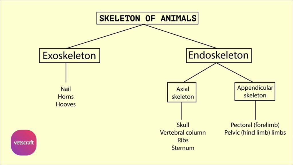
The sacrum is a part of the vertebral column in domestic animals, formed by the fusion of multiple sacral vertebrae. It serves as a strong, wedge-shaped structure that connects the spine to the pelvis and supports the hind limbs.
Sacrum of Ox
The sacrum of ox is formed by the fusion of five sacral vertebrae. It is triangular in form and is wedged in between the ilia, with which it articulates on each side. Its long axis is strongly curved, and it presents two surfaces, two borders, a base, and an apex.
The dorsal surface is widest in front and shows, along its median line, a crest—the median sacral crest—which is formed by the fusion of spinous processes of the five sacral vertebrae. The summit of this crest is rough and tuberous.
On either side of this crest is another crest—the lateral sacral crest—which results from the fusion of articular processes. The dorsal surface shows four dorsal sacral foramina for the passage of the dorsal primary branches of the sacral spinal nerves.
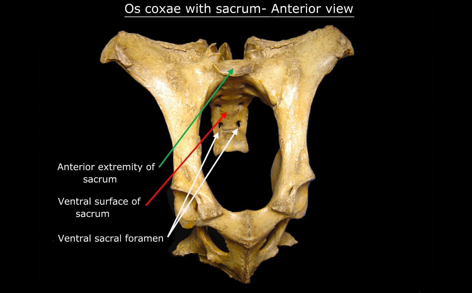
The ventral surface is arched and forms the roof of the pelvis. This surface is marked centrally by a faint longitudinal groove for the middle sacral artery and also by four transverse lines, which indicate the lines of fusion of the bodies of the sacral vertebrae. There are four large ventral sacral foramina at the ends of the transverse lines on either side for the ventral primary branches of the sacral spinal nerves.
The lateral borders are thin, concave, and serve for the attachment of the sacro-sciatic ligament.
The base or anterior extremity of the sacrum is, in fact, the anterior end of the first sacral vertebra. It shows, centrally, the body of the first sacral segment, which is wide, flattened from above downwards, and elongated transversely. It articulates with the posterior end of the body of the sixth lumbar vertebra, and the ventral margin projects slightly, forming the promontory. The edge of the pedicles shows the usual anterior notches, which, with the posterior notches of the last lumbar, form the intervertebral foramina for the sixth lumbar spinal nerves.
The anterior part of the sacral (vertebral) canal is triangular, with its angles rounded off. Traced backward, its size diminishes, and the posterior opening is small and triangular. On either side of the arch and above the notches are the anterior articular processes, which are very large and widely separated.
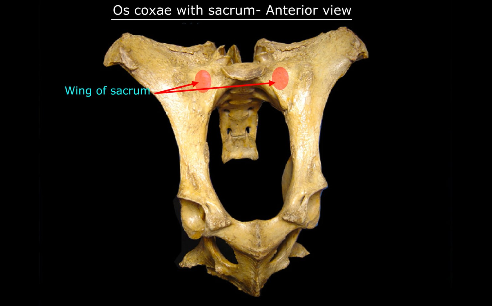
The lateral parts of the base are the alae or wings. Each wing is a quadrilateral compressed plate directed downwards and forwards. The anterior face is extensive, concave from side to side, and non-articular. The posterior face is rough, and in its lower part there is a triangular area—the articular surface for articulation with the ilium.
The apex or posterior extremity of the sacrum is the posterior end of the fifth sacral vertebra. Centrally, it presents a small triangular neural ring and the posterior notches. Above these is the posterior extremity of the median sacral crest. Placed below the neural ring is the centrum of the last sacral segment, which is flat.
Comparative Anatomy of Sacrum
The sacrum exhibits notable anatomical differences across domestic animal species. While it is generally composed of fused sacral vertebrae in all species, the number of segments, degree of fusion, orientation of wings (alae), spinal canal shape, and articular surfaces vary significantly.
Sheep and Goat
- The sacrum of sheep and goats consists of four segments ordinarily, but the last vertebra may remain separate or undergo partial fusion.
- There is no vascular groove on the ventral surface.
- The dorsal spines are not fused, with the exception of the first two.
- The transverse process of the last segment is distinct.
Horse
- The sacrum presents five separate dorsal spines, the bases of which are fused in the old horse.
- On either side of the bases of the spines is a groove in which are the four dorsal foramina.
- The ventral surface is not as deeply arched as in the ox.
- The wings are prismatic with pointed ends. Each has a large oval convex facet in front, which articulates with the facet on the posterior border of the transverse process of the last lumbar vertebra.
Pig
- Usually, four sacral vertebrae are present in pigs.
- They fuse in the later part of life.
- The spines are little developed.
- The anterior articular processes are very large.
Dog
- The sacrum of the dog is formed by three segments and is short, wide, and quadrangular.
- The articular processes are vestigial and are represented on the side of the median crest as a pair of tubercles, and lateral to these are the two dorsal sacral foramina.
- The pelvic face is deeply concave and presents two ventral sacral foramina.
- The wings are prismatic and very high. Their lateral faces are extensive, face almost directly inward, and bear an auricular surface on the lower part.
- The promontory is distinct.
- The transverse processes of the last segment project backward and may articulate or fuse with those of the first coccygeal.
- The spinal canal is compressed dorso-ventrally.
Rabbit
- The sacrum of the rabbit is a single bone in the adult animal, formed by the fusion of four vertebrae.
- The articulation with the pelvic girdle is primarily on the transverse process of the first sacral vertebra.
Fowl
- The lumbar and sacrum are fused and called lumbosacrals in fowl.
- They are fourteen in number, and these, with the seventh dorsal and the first coccygeal—altogether 16—are fused into one mass called the synsacrum. This forms a rhomboid mass included between the two pelvic bones.
- The spines form a crest in the anterior third but are absent posteriorly.
- The transverse ridges on the ventral face indicate the positions of the transverse processes. The extremities of all the transverse processes fuse with the medial border of the ilium.