Rabies (Mad Dog Disease)
Rabies (Mad Dog Disease) is acute viral encephalitis of all warm-blooded animals characterized by altered behaviour, aggressiveness, progressive paralysis and in most species by death.
Rabies (Mad Dog Disease) is also known as Hydrophobia and Lyssa in animals.
Rabies (Mad Dog Disease) is an acute progressive viral encephalomyelitis that primarily affects carnivores and bats, although it can affect any mammals. The disease is fatal once the clinical signs are appear.
Etiology
- Rabies is caused by Lyssa virus of family Rhabdoviridae.
- It appears as filamentous, bell or bullet shaped and approximately 60-175 nm in size. The virion is rounded at one end and flat or truncated at the other.
- It is readily inactivated by sunlight, drying in air 40-70% alcohol, quaternary ammonium compounds, tincture-iodine, carbolic acid and any lipid solvents.
- The virus is very resistant to autolysis and putrefaction. The virus will persist for months in infected nervous tissue in 50% glycerol. It can be preserved at –700C or by lyophilization.
Epidemiology
- At the present time rabies occurs in most parts of the world except in Japan, UK, New Zealand, Antarctica, Australia, Hawaii islands and Switzerland. In India the incidence is very high.
Host affected
- Fox, wolf, coyotes and jackal are extremely susceptible.
- Guinea pig, Hamster, Bat, Mongoose, mice, rabbit, skunk and cattle are highly susceptible.
- Dog, sheep, goat, horse and human are moderately susceptible.
- Poultry and opossum are resistant.
- Reservoir animal for rabies vary throughout the world.
Transmission
- Introduction of virus laden saliva into tissue usually by the bite of rabid animal.
- Saliva from salivary gland or brain less likely to cause infection by entering the body through fresh wounds or intact mucous membrane.
- Hematogenous spread does not occur.
- Aerosol transmission is not dangerous in most circumstances, however, aerosol transmission has occurred under very specialized conditions in which high concentration of virus in suspended particles.
Pathogenesis
- Following introduction of virus into tissue, the virus travels via peripheral nerves to the spinal cord and ascends to the brain, replication in brain causes neuronal damage.
- The virus spread centrifugally within nerve cell processes and is released at axonal terminal, where it infect the salivary glands.
- The virus sheds in saliva intermittently.
Clinical manifestation
- The incubation period in natural outbreak of dog rabies is 3-8 weeks, but it may be as short as 10 days to as long as one year.
- The clinical signs of rabies is rarely definitive.
- The most reliable signs regardless of species, are acute behavioural changes and unexplained progressive paralysis.
- Behavioural changes may include sudden onset of anorexia, signs of apprehension or nervousness, irritability and hyper excitability, altered phonation and changes in temperament.
- Uncharacteristic aggressiveness- normally docile animal may suddenly become vicious.
- The clinical course may be divided into three general phases.
- Prodromal, acute excitative, and paralytic end stage.
Prodromal phase
- Affected animals are often confused and disoriented.
- Wild animal may lose their natural fear of humans.
Furious form
- The animal becomes irritable, and with slightest provocation aggressively use its teeth, claws, horns or hooves, alertness and anxiety.
- Noise may invite attack.
- Attacking other animals.
- Eating inanimate object.
- Papillary dilation.
Paralytic form
- It manifest as ataxia, paralysis of throat and masseter muscles often with profuse salivation and inability to swallow.
- Dropping of jaw.
- Paralysis progress rapidly to all parts of the body, coma and death in few hours.
Cattle
- Abnormal bellowing, which may continue or intermittently until before death.
- Drooling of saliva.
- The eye and ear follows sound and movement.
- Yawning movements are more accurately described as voiceless attempt to bellow.
- In paralytic form, knuckling of the hind fetlock, sagging and swaying of the hindquarters while walking, deviation or flaccidity of tail to one side.
- Decreased sensation usually accompanied this weakness.
- Tenesmus with paralysis of anus, resulting in the suckling in and blowing out of air.
Sheep
- The clinical symptoms are similar to cattle, some animals show sexual excitement, attacking human, wool pulling, sudden falling after violent exertion, muscle tremor and salivation.
- Excessive bleating does not occur.
Goat
- Goats are aggressive and continuously bleating.
Horse
- Evidence of distress, extreme agitation, these signs may accompanied by rolling.
- Horse may bite or strike viciously.
- Animal frequently have self implicated wound.
- Muzzle tremors and pharyngeal paresis are common.
- Sudden onset of lameness in one limb followed by recumbency, high stepping gait, blindness, paddling, convulsions and terminally paralysis.
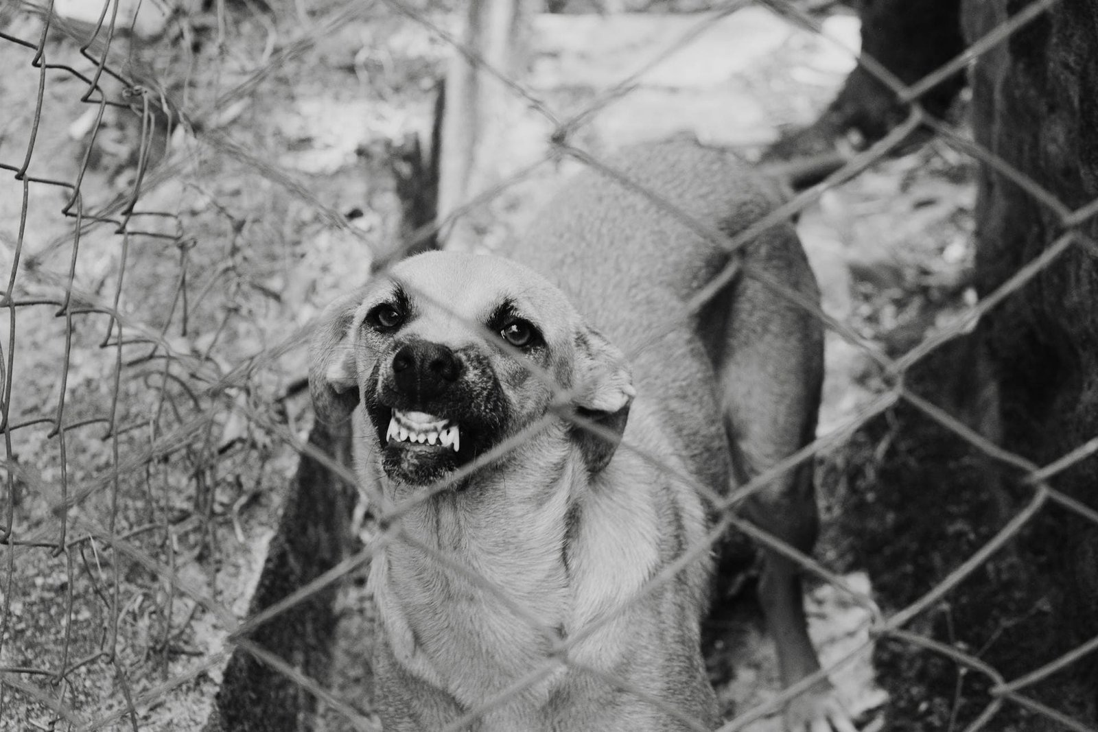
Dog
- Dog becomes irritable, restless, nervous, deprived appetite, aggressive, and often dangerous as it loses all fear of humans and bites at anything that gains its attention.
- There is usually exaggerated response to light and sound.
- Dog exhibit characteristic change in its barking and may howl in an unusual tone due to the paralysis of laryngeal muscle.
- Salivation and frothing at the mouth becomes progressively more profound.
- Terminally, there are often convulsive seizures, coma and respiratory arrest, with death occurring 2 to 14 days after the onset of clinical signs.
Cat
- In cats the clinical signs are similar to those of dogs but lost for 2-4 days before death occur.
Pig
- Tendency to attack, twitching of the nose, rapid chewing movements, excessive salivation, walk backward and terminally paralysis.
Sample collection
- Live animals-Saliva, corneal/conjunctiva smear.
- Dead animals- Hippocampus major, cerebellum, cerebral cortex and placed in 50% glycerol saline to preserve the virus.
- No refrigeration is required.
Diagnosis
- Based on clinical signs.
- Demonstration of Negri bodies in impression smear collected from hippocampus major in dog and cerebellum in cattle by sellers staining.
- Negribodies are intracytoplasmic, magenta red or cherry red spherical or oval bodies with characteristic basophilic inner granules in the cytoplasm of neurons.
- Animal inoculation: For isolation of virus, the brain or other tissue specimens are prepared as 10% suspension.
- Suckling mice or hamsters of 3-6 weeks age are generally used for isolation and identification.
- Route of inoculation is intracerebral. Infected mice almost always develop clinical symptoms within 17 days of inoculation.
- Direct fluorescent antibody test (FAT) on acetone fixed brain tissue smears. It is gold standard test for diagnosis of rabies.
- Rapid fluorescent focus inhibition test (RFFIT) rarely used to confirm diagnosis due to late seroconversion.
Differential diagnosis
- Nervous ketosis
- Polioenchalomalacia
- Lead poisoning
- Lactation tetany
- Vitamin A deficiency
- Pseudorabies
- Enterotoxaemia
Treatment
- Supportive care for rabies infected animal is not recommended.
Prevention and control
- For control of rabies in dog population, vaccination of minimum 70% of dog should be necessary. Read more (Prevention and control).

