Pterygoid Bone of Domestic Animals: Anatomy of Ox, Horse, Dog and Fowl
The pterygoid bone is an important paired bone of the skull in domestic animals. It contributes to the formation of the posterior nasal cavity, supports the soft palate region, and participates in the formation of the pterygoid canal. The shape and articulations of the pterygoid bone vary among species such as ox, horse, dog, and fowl.
Pterygoid Bone of Ox
The pterygoid bones of the ox are thin bony plates situated on either side of the posterior nares, overlying the perpendicular parts of the palatine bones.
The medial surface forms part of the lateral wall of the posterior nares. The lateral surface articulates with the palatine bone and the subsphenoidal process of the sphenoid bone.
The extreme upper part of this surface forms the pterygoid (Vidian) canal together with the sphenoid bone.
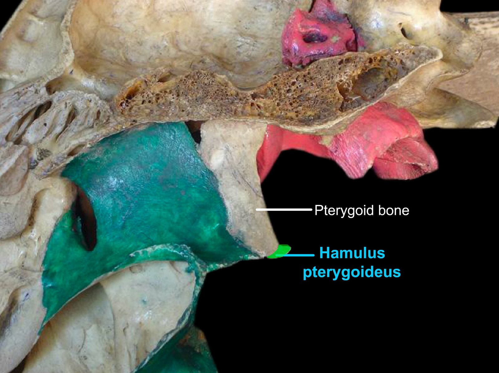
The lower extremity is free and forms a curved process, the hamulus pterygoideus, around which the tendon of the tensor veli palatini muscle is reflected.
Pterygoid Bone of Horse
The pterygoid bone of the horse is shorter and narrower. The lateral surface articulates with the palatine, vomer, and sphenoid bones, contributing with the latter to the formation of the pterygoid canal.
Pterygoid Bone of Dog
The pterygoid bone of the dog is short, broad, and quadrilateral in shape.
Pterygoid Bone of Fowl
The pterygoid bone of the fowl is rod-shaped and extends between the rostrum of the sphenoid medially and the quadrate bone laterally.