TABLE OF CONTENTS
Pregnancy Diagnosis in Cows
Pregnancy diagnosis (Cyesiognosis) in cows done by various methods like external appearance, rectal examination and palpation, laboratory methods, fetal echocardiography, etc.
Accurate early pregnancy diagnosis in cows is essential for successful breeding programme.
Ability is required for successful practice. Skill is to be developed to indicate the duration within weeks in early and months in late pregnancy.
External Indications of Pregnancy
- Cessation of estrum-under field conditions-hardly an useful information.
- Increase in abdominal size-not reliable.
- Enlargement and edema of udder-in heifers-begins around 5 months-older cows last 1-4 weeks.
- Relaxation of pelvic ligaments and edema and relaxation of vulva in last weeks.
Rectal Examination for Pregnancy Diagnosis
Pregnancy diagnosis is based on the following:
- Presence of the amniotic vesicle
- Increase in size of the uterus or dissimilarity of horns
- Fluid feeling and thinning of the uterus
- Slipping of the fetal membranes
- Feel of the fetus and its bump
- Feel of the cotyledons
- Enlargement of the middle uterine artery
- Presence of Corpus luteum (CL)
Above signs are present in all pregnancies. However, certain signs alone are not enough to declare an animal as pregnant.
Positive Signs of Pregnancy
- Amniotic vesicle
- Fetal membrane slip
- Fetus
- Cotyledons
Without confirming anyone of these positive signs no cow should be declared pregnant.
Uterine Changes During Pregnancy
- The gravid horn and to a lesser extent the nongravid horn increase in size because of the growing fetus and fetal fluid-leading to an enlargement and dissimilarity in size between the horns.
- The increase in size leads to thinning of the uterine wall giving a characteristic fluid, watery “alive” feeling.
- From 40–90 days the uterus feels somewhat like a thick rubber balloon nearly filled with water.
- The uterus remains mostly in the pelvic cavity upto 3 months afterwards descends into the abdominal cavity.
Amniotic Vesicle
- Palpable in the free part of the uterine horn from about 30 days because of its tense spherical nature feeling like a soft shelled hen‘s egg, and gently palpated along their entire length between the thumb and middle two fingers. The amniotic sac can be felt as a distinct round or oval turgid object (like a soft shelled hen‘s egg) slipping between the fingers.
- The sac should not be compressed directly but gently pushed backwards and forwards.
- Usually be located in the pregnant horn in the vicinity of fetal fluid enlargement and thinning of uterine walls.
Fetal Membrane Slip
Fetal membrane slip best performed between 30 and 90 days of pregnancy of cows.
Method of Fetal Membrane Slip
- Identify the bifurcation of uterine horns.
- Pick up and pinch or compress the enlarged gravid horn between the thumb and either the index or middle finger just cranial to the bifurcation.
- Allow the structures to slip and identify the slipping of the fetal membrane first and then the uterine wall (Double slipping).
- In very early pregnancy-it is recommended to grasp the entire horn and let it slip on the ventral aspect so that the thicker connective tissue band can be easily recognized.
- After 70 days of gestation it is often easier to slip in the nongravid horn.
Fetus
- Palpation of the fetus before 60-70 days is not possible because of the smaller size and tenseness of the amniotic sac.
- Rocking of the flat hand over the enlarged uterus sets the fetal fluids in motion and results in rebounce of the fetus against the hand (fetal bump). Upto 4-5 months the fetal bump can be easily made.
- In late gestation after 8 months fetal parts (not bump) are easily palpable.
Placentomes
- Become recognisable by rectal palpation at about 90–100 days.
- First felt in the mid line by pressing down the uterine body and the base of horns.
- In early stages it is difficult to identify them as distinct individual structures.
- The uterus feels as if it has an irregular corrugated surface and feels like a sack full of small potatoes.
- Once the uterus sunk into the abdomen between 5 and 7 months it is frequently impossible to palpate them.
Hypertrophy and Fremitus of the Middle Arteries
- In non-gravid and early pregnancy-impossible to identify
- The artery runs in the broad ligament along a tortuous course passing downwards and forwards over the pelvic brim.
- Inexperienced persons sometime confuse it with iliac and obturator arteries which are highly fastened.
- Middle uterine artery is very mobile and can be encircled with the thumb and finger.
- The characteristic hypertrophy and “whirring” (feels like water surging intermittently through a thin rubber hose) is noted in pregnant cows.
Palpation at Different Stages
35–40 days of Pregnancy
- Requires more skill.
- Easily detectable in heifers and difficult in older cows.
- Uterus in the pelvic cavity.
- Slight enlargement on the free part on one horn with fluid feeling.
- Detectable dorsal bulging.
- Amniotic sac about the size of the yolk of a hen‘s egg.
- CL ipsilateral to the horn containing amnion.
45–50 days of Pregnancy
- Uterus in the pelvic cavity.
- Difference in size of pregnant and nonpregnant horn.
- Dorsal bulging more pronounced.
- Amnion about the size of a small hen‘s egg (soft shelled egg).
- Membrane slip.
60 days of Pregnancy
- Uterus in the pelvic cavity
- Uterus feels like a narrow balloon filled with water.
- Dissimilarity of horns.
- Membrane slip in both horns
- Amnion not detectable
- Diameter of the gravid horn is about 6-9 cm.
90 days of Pregnancy
- The uterus may be still in the pelvic cavity or over the pelvic brim.
- Uterus about the size of a football bladder.
- Dissimilarity of horns.
- Fetal bump easily palpable.
- Diameter of the gravid horn is about 10-13 cm.
120 days of Pregnancy
- The uterus descends into the abdomen.
- Uterine contour is still palpable.
- Fetus can be palpated.
- Small placentomes can be identified.
- Fremitus detected.
- Diameter of the gravid horn is about 12.5-18 cm.
150 days of Pregnancy
- Uterus in the abdominal cavity.
- The cervix over the pelvic brim.
- Distinct crowded placentomes about the size of ovaries palpable.
- Fetus may or may not be palpable.
- Fermitus quite distinct.
170–230 days of Pregnancy
- Cervix at the brim of the pelvis.
- Tight cord like folds of uterus running deep into the abdomen from the cervix.
- Uterus difficult to palpate.
- Placentomes and fetus also difficult to palpate.
- Very distinct fermitus.
230–280 days of Pregnancy
- The fetus extends back towards the pelvic cavity.
- The head and front feet palpable.
- Movement of fetus reflex can be detected.
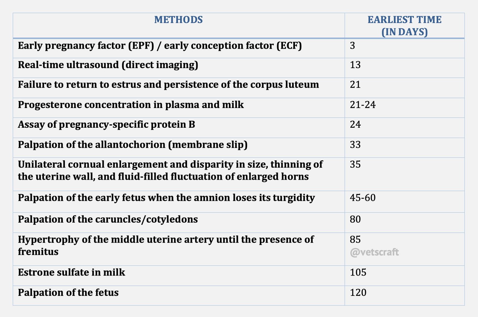
Laboratory Methods of Pregnancy Diagnosis
Laboratory Methods of Pregnancy Diagnosis can be done by various methods:
Hormone Estimation:
- Progesterone in milk and plasma
- Estrone sulfate in milk
By Proteins:
- Bovine Pregnancy Specific Protein-B.
- Immunosuppressive Early Pregnancy Factor.
Progesterone in Milk and Plasma
- For diagnosing pregnancy by determination of progesterone concentration in the plasma of cows, using Radioimmunoassay (RIA).
- P4 concentrations in blood and milk are elevated at 20-24 days post insemination. Conversely, if pregnancy fails, P4 concentration is elevated until approximately day 17 when luteolysis is followed by a sharp decline in P4 concentration by day 20 and return to estrus.
- Optimum time for collecting the milk sample: 24 days after breeding.
Estrone Sulphate in Milk
- Estrone sulfate is a product of the placenta and is present in the milk of pregnant cows in concentrations sufficient to differentiate between the pregnant and non-pregnant cows after approximately day 100 of gestation.
- Practically, however, assays for estrone sulfate are not useful for early detection of pregnancy and offer no substantial advantage over other methods except in the case of a few cows in which rectal palpation cannot be performed.
Bovine Pregnancy Specific Protein-B
- In bovines a pregnancy-specific protein (bPSPB) secreted by the trophoblastic cells has been isolated and purified.
- Concentrations of bPSPB are detectable in a few cases as early as 15 days after insemination and in nearly all pregnant cows by 24 days after insemination.
Immune Suppressive Early Pregnancy Factor
- Blood serum assayed within 24 hrs of ovulation for the presence of immunosuppressive early pregnancy factor.
- The assay is able to diagnose pregnancy in 87.5% cows at less than 24 hours of gestation and 12.5% inaccurate in the identification of non-pregnant cows.
Ultrasonography (USG)
- In animals, transducers of 5 MHz and 7.5 MHz frequencies are most widely used for transrectal ultrasonography.
- Under practical conditions, 5 MHz transducer is an accurate method for pregnancy diagnosis after approximately day 24. A 3.5 MHz transducer is found to be reliable after day 30.
Fetal Echocardiography
Fetal Echocardiography is not applicable before 5 months of gestation, but might have application for the diagnosis of multiple pregnancies. Refer to practical module on Ultrasonography for further details on instuments, technique etc.
Ultrasonographic Observations
- The embryo proper is first detected within the amniotic vesicle on day 20, when it is 3.5 mm length. By day 60, the embryo grows to 66.1 mm.
- Between days 28-31, fore limb buds become visible and hind limb buds approximately 2 days later.
- Two claws become visible on the hooves between days 42-49.
- Movements of the fetal head and feet are first detected between days 42-50.
- Ribs could be visualized beginning on days 51-55.
- Placentomes are first visualized between days 33-38 in the area of the embryo and then can be seen throughout the uterine horn by day 60.
Prostaglandin Induced Milk Flow Test
Diagnosis of pregnancy in cows based on the observation of milk ejection which in the case of CL maintenance results from the release of luteal oxytocin induced by intravenous administration of a non-luteolytic dose of PGF2 alpha.
- Examine all cows per rectum on day 18 post insemination to assess ovarian status.
- Perform PG-IMF test on the same day 3 hours after evening milking.
Preparation of Non-Luteolytic Dose of PGF2 alpha: One ml of PGF2 alpha (lutalyse) which contains 5000 micro gram is reconstituted in 39 ml of distilled water to arrive at a final concentration of 125 micro gram/ml.
Protocol for this test
- After washing the udder and teat, a sterile cannula is placed in the left fore teat to empty the cistern milk.
- Subsequently a non-luteolytic dose of 125 micro gram of PGF2 alpha is injected through the ear vein.
- After the injection the time duration of milk flow is recorded.
Inference
Elicitation of milk let down reflex with free flow of alveolar milk within a few seconds after injection and lasting for 3-5 minutes is considered to have a functional CL/presence of conceptus.
Absence of milk flow is indicative of non-functional CL.
The prostaglandin induced milk let down response observed in pregnant animals could be attributed to the release of endogenous luteal oxytocin that was actively synthesised by the luteal tissue and was available for immediate release in response to PGF2 alpha resulting in alveolar milk ejection immediately by increasing the intramammary pressure within the cistern and enlargement of cisternal volume.
Differential Diagnosis
Uterine enlargement is usually associated with pregnancy. It should not be always construed that it is the only cause.
The ability to make an accurate, early diagnosis is required of most successful large animal practitioners. Hence, it is imperative to differentiate physiological uterine enlargement (gravid) at each stage of pregnancy from that of one or more of the other causes.
While performing a rectal palpation to diagnose pregnancy, a careful consideration of anatomical structure and relationships of the organs and their consistency, will help to prevent erroneous diagnoses.
Anatomical structures to be differentiated:
- Distended urinary bladder
- Pendulous left kidney
- Rumen


