TABLE OF CONTENTS
Postpartum Uterine Infections in Animals
Postpartum uterine infections in animals are bacterial infections that develop in the uterus following parturition.
They occur due to contamination of the uterine environment when the cervix remains open after calving, particularly if there has been dystocia, retained placenta, poor hygiene, or trauma to the reproductive tract.
Types of Infection in Uterus
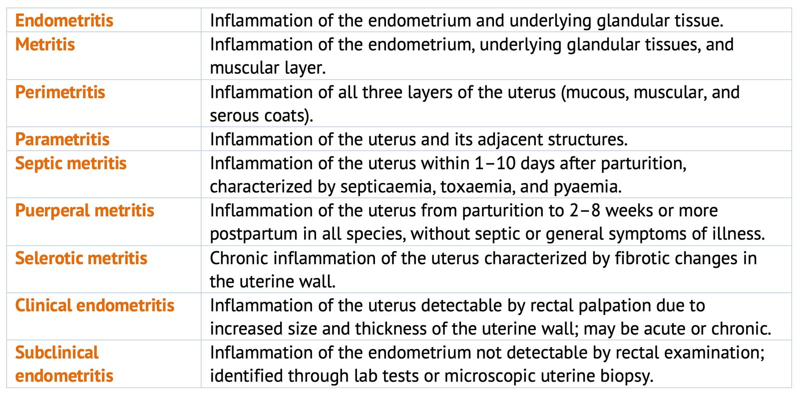
- Endometritis: Means inflammation of the endometrium and underlying glandular tissue.
- Metritis: Inflammation of the endometrium, underlying glandular tissues and muscular layer.
- Perimetritis: Inflammation of all the three layers of the uterus (mucous, muscular and serous coats).
- Parametritis: Inflammation of uterus and its adjacent structures.
- Septic metritis: Inflammation of uterus within 1–10 days after parturition. It is characterized by septicaemia, toxaemia and pyaemia.
- Puerperal metritis: Inflammation of uterus from the time of parturition to 2 to 8 weeks or more postpartum in all species. It is not accompanied by septic or general symptoms of illness.
- Selerotic metritis: Chronic inflammation of the uterus characterized by fibrotic changes in the uterine wall.
- Clinical endometritis: Inflammation of the uterus can be detected by rectal palpation as an increased in size and thickness of the uterine wall. It may be acute or chronic in nature.
- Subclinical endometritis: Inflammation of the endometrium cannot be detected by rectal examination. It can be detected by laboratory examination or microscopic examination of uterine biopsy.
Postpartum uterine infections in animals are usually associated with uterine atony or inertia. The more pathogenic types of organism present in the uterus and their toxins are absorbed into the circulation producing several general symptoms associated with septicaemia, toxaemia and pyaemia.
Etiology
The uterus is normally protected from bacterial contamination by the vulva, vestibular sphincter and cervix.
Immediately after parturition these mechanical barriers are breached and the uterus is contaminated by a variety of pathogenic and non pathogenic organisms.
Most of these bacteria are only transient residents and are promptly eliminated by the uterine defence mechanism during the puerperial period.
In some cases these pathogens persist in the uterus and causes diseases.
Bacteriology
The most commonly associated organism is C. Pyogenes. Gram negative anaeraobes like Fusobacterium and Bacteroides are frequently associated with C. Pyogenes. These organism decrease chemotaxis and inhibits phagocytosis by neutrophils. Clostridium spp. occasionally invades the uterus and cause gangrenous metritis.
These infections in cows resulted in lochia assuming a white, yellow white or grey mucopurulent character towards the later part of the puerperal period. The bacterial invasion coincides with the increasing amount of lochia, which together with the intrauterine temperature and pH create a suitable environment for bacterial multiplication.
Major Groups of Uterine Bacteria
Coliform Bacteria
- E. coli
- Proteus spp.
- Enterobacter spp.
Incidental Bacteria
- Streptococci spp.
- Staphylocci spp.
- Pasteurella spp.
- Bacillus spp.
Corynebacteria
- C. pyogenes
Gram Negative Anaerobic Bacteria
- Bacteroides spp.
- Fusobacterium spp.
Gram Positive Anaerobic Bacteria
- Clostridium perfringes
- C. Sporogenes
- Other clostridium spp.
Unsanitary Calving Conditions
If the calving environment is dusty or wet or is often used without sanitation pathogenic bacteria increased in numbers.
Other Periparturient Diseases
Retained fetal membranes are associated with increased puerperal metritis. Consequently periparturient conditions that increase retained fetal membranes also increase postpartum uterine disease (e.g. Vitamin A or Selenium deficiencies, excessive weight gain during the dry period, multiple birth, heat stress, short gestation, abortion, still birth, dystocia).
Endocrine Factors
Concentration of low estrogen (<100 pg/ml) or of high progesterone (>7–9 ng/ml) alone are associated with increase in the incidence of retained fetal membranes.
Poorly Selected Uterine Treatment
Many of the antimicrobial and disinfectants infused into the bovine uterus lower the uterine defence mechanism and irritate and damage the endometrium. Irritating substances in the oviduct may also result in tubal inflammation and perimetritis.
Phagocytosis of microorganisms and Neutrophills is important in the bovine uterine defence system. This defence system is stimulated about 2 days postpartum by invading microorganisms. When uterine defence mechanism is impaired under pathologic condition such as delivery of dead or weak calves, Phagocytosis can be depressed for several days to several weeks. Trauma to the genital tract by removal of retained fetal membranes and by obstetrical procedures also depress Phagocytosis.
Nutritional Aspect
Metritis and retained fetal membranes are more prevalent in overfed cows. Improper ratio of calcium to phosphorus fed prior to calving and defieiencies of Vitamin A and Vitamin E and selenium increases the incidence of metritis and retained placenta. Debilitated cows are probably less resistance to puerperal infections.
Uterine Defence Mechanism
- Normally post partum uterus has a heavy load of bacteria, which gets eliminated by defence mechanism (stimulated about 2 days after parturition) include Phagocytosis by neutrophils.
- Under pathological condition such as abnormal parturition (dead calf, Trauma to genital tract by removal of RFM and obstetrical procedures etc) the Phagocytosis is compermised for several days to weeks.
- Possibly the number of antibiotics present in uterus might also depress the defence mechanisms.
- Uterine defence mechanisms is stimulated by estrogen and inhibited progesterone.

