TABLE OF CONTENTS
Postpartum Haemorrhage in Animals: Causes, Symptoms, and Treatment
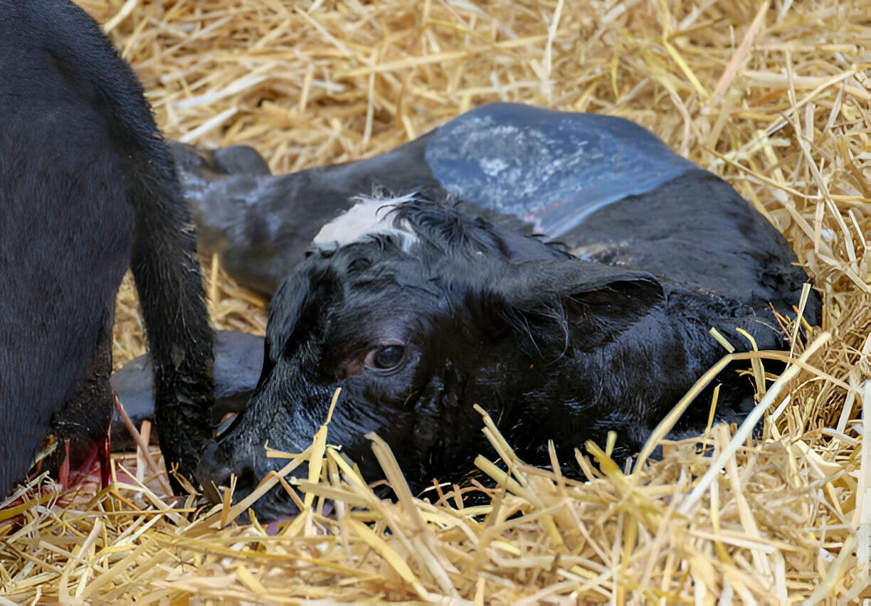
Postpartum haemorrhage is defined as abnormal or excessive bleeding from the reproductive tract occurring after the expulsion of the fetus and placenta.
Postpartum haemorrhage, or bleeding into the uterus or birth canal, may occur after parturition due to trauma, lacerations, or rupture of the genital organs.
Postpartum Haemorrhage in Livestock
Etiology
Haemorrhage in the uterus may be due to bleeding from an incised caruncle or caruncular stalk in the cow or ewe, from the incised or lacerated endometrium in the mare, or from premature removal of the fetal membranes or placenta.
This accident may occur at the time of parturition, during a caesarean section or fetotomy, or may take place later due to improper or too early removal of a retained placenta in any uniparous animal.
Symptoms
Severe intrauterine hemorrhage may occur from several hours to a day or so after calving and drooping of the placenta that results in a massive blood clot filling the gravid horn of the uterus.
Rarely, death may occur.
Slight bleeding may be observed from the ruptured end of the umbilical cord or from slight lacerations of the uterus, cervix, vagina, or vulva.
In severe lacerations or rupture—particularly in the cervix, vagina, and in rare instances, the vulva—hemorrhage may be profuse due to a rupture of a large vessel.
Blood may flow in a stream from the vulva as soon as the fetus is removed. Most of these lacerations and injuries follow forced extraction.
Intraperitoneal or intrapelvic hemorrhage may occur and, if severe enough, produce acute symptoms of anemia and rarely death, especially in the mare. This is ordinarily seen in cases of dystocia, rupture of the uterus and uterine vessels before, during, or after correction of torsion of the uterus, and in trauma, especially in fetotomy operations or forced extraction in young heifers.
These hemorrhagic conditions would be greatly aggravated in cattle fed sweet clover.
Treatment
In the treatment or handling of these conditions the usual surgical procedures to control the haemorrhage and supportive treatment are indicated.
Most cases of slight bleeding from the genital tract at the time of parturition are not serious and require n o treatment.
In severe cases, bleeding from the uterus of large animal’s posterior pituitary hormone or oxytocin, 20 to 25 units, may help to control haemorrhage by contracting the uterus and its vessels.
Injecting 500 ml of saline to which 10 ml of formalin has been added, or 500 ml of calcium borogluconate may also aid in hastening the clotting of blood, and thus control the haemorrhage.
If bleeding occurs from a large vessel through a laceration in the vaginal wall, the vessel may be clamped by forceps. These should be left in place for 24 to 48 hours and then may be broken down manually the next 2 or 3 days until entirely removed.
If the clot should not be discovered for several weeks or months the uterus would probably break it down and absorb it. Injections of estrogens as in cases of mummification of the fetus may aid the expulsion and absorption of the clotted blood.
Intra pelvic or perivaginal bleeding may cause a stenosis of the vagina during or after forced extraction or fetotomy by neither it nor the intrauterine hemorrhage is usually fatal.
In 12 to 21 years old mares rupture of the uterine vessels and sudden death due to hemorrhagic shock may occur before, during or after an apparently normal gestation and parturition.
The middle uterine artery was most commonly involved but the iliac or utero–ovarian arteries occasionally were affected.
Most fatal haemorrhages occur intraperitoneally due to rupture of the large vessels in the broad ligament caused by degenerative changes in the vessel wall, especially in horses, or by torsion of the uterus or prolapse of the uterus.
Definite symptoms of severe hemorrhage may be observed, indicated by weakness, depression, very rapid pulse and respiration rates and pale mucous membranes.
If the operator promptly enters the peritoneal cavity through the abdominal wall in the flank region or through the uterine wall in prolapsed, he may be able to control bleeding by ligating the ruptured vessels.
The prognosis is very poor in these cases as severe hemorrhage, shock and death may occur in rapid succession.
Postpartum Haemorrhage in Mares
In mares early signs of colic, sweating, pain, rapid pulse rate and moderate anemia may occur due to rupture of a uterine vessel with relatively slow loss of blood between the two layers of the broad ligament causing a large haematoma.
If this ruptures intraperitoneally, then severe acute signs of shock, rapid weak pulse, anemia, prostration, and death follow. If hemorrhage is severe enough to cause clinical symptoms, blood transfusions of 200 to 8000 ml or more in large animals, saline injections, gelatin, or other types of solutions designed to maintain blood pressure should be given and repeated as often as necessary.
Excessive fluids should be avoided. The mare should be sedated with a large dose of tranquilizer and closely confined.
In lacerations or ruptures of the genital tract preventive treatments should be used to control the infection, such as the administration of sulfanomides, antibiotics, or local mild antiseptics.
Thrombosis of the large uterine arteries and veins is occasionally observed.
Thrombi are seen most often in the veins following prolapse of the uterus or uterine torsion when circulation has been restricted.
Occasionally an aneurysm of the middle uterine artery may be palpated on rectal examination after parturition in the cow, or a haematoma in the broad ligament of the mare.
Adhesions between the genital tract and ovaries and other pelvic and abdominal organs and tissues may occur following postpartum haemorrhage.

