TABLE OF CONTENTS
Ovulation in Animals
Ovulation in animals is the release of oocyte from mature Graafian follicle. Ovulation in mammalian ovary occurs on any point in the ovarian surface while ovulation in mares is restricted to ovulatory fossa.
In cow, sheep and horses, ovulation occurs at random irrespective of which ovary contains previous corpus luteum (CL). However, in some animals ovulation consistently alternates between ovaries and in others (whales) ovulation may predominate in one ovary.
In the rhesus monkey, the CL retards subsequent follicular growth so that ovulation alternates between ovaries.
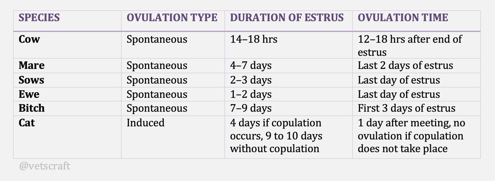
There are two types of ovulators:
- Spontaneous ovulators (eg. Cow, mare, sheep, goat, dog). Ovulation occurs spontaneously irrespective of whether mating has taken place or not.
- Induced ovulators or Reflex ovulators (eg. Cat, Mink, Ferret). Copulation is a must for ovulation to take place.
Theories of Ovulation
There are various theories of ovulation in animals:
- Follicular pressure theory
- High osmotic pressure theory
- Ischemic theory
- Follicular wall thinning and rupture
(1) Follicular pressure theory
As the follicle grows the amount of liquor folliculi also increases. This liquor folliculi exerts pressure on the follicular wall thereby follicles rupture. However, this was not the case with cystic ovaries where there was both an increase in follicular size and follicular fluid but there was no rupture. Theory not accepted.
(2) High osmotic pressure theory
Liquor folliculi consists of more amount of electrolytes particularly Na and K. increased osmotic pressure leads to rupture of the follicle. in cystic ovaries size of the follicle and amount of follicular fluids along with electrolyte content also increased but still there was no rupture. Hence, theory was disagreed.
(3) Ischemic theory
Increased follicular fluid exerts pressure against follicular wall. At one point due to pressure, ischemia occurs and leads to stigma formation and ovulation. Theory partly accepted.
(4) Follicular wall thinning and rupture
Nearing ovulation, the blood supply to the follicle increases. Thinning of the follicular wall occurs at one point called as stigma. Ovarian contractions and follicular rupture occurs. Theory accepted.
Recent theory is that ovulation is a combination of physiological, biochemical and biophysical mechanisms.
A surge of LH occurs at the beginning of oestrus prior to ovulation when progesterone is at its minimal levels and oestradiol has reached its highest cyclical values.
Several tissue layers separate the oocyte from the outside of the follicle. these are the surfaced epithelium, the tunica albugenia, techa externa and interna, basement membrane and granulose cells. All these tissue layers have to be broken down before ovulation can take place.
An increased blood flow near ovulation occurs to all classes of follicles but the follicle destined to ovulate receives the largest volume of blood and has capillaries more permeable then those in other follicle. As the follicle enlarges it begins to protrude from the surface of the ovaries, the vascularity of the follicular surface increases except at its centre, which is devoid of blood vessels. This avascular area is the future point of rupture. Meanwhile there is dissociation of cumulus cells which detaches the oocyte from the membrane granulose and now the oocyte is surrounded by the radiate cells. Resumption of meiosis (Nuclear Maturation) occurs 3 hours after LH surge and ends 1 hour before ovulation when the first polar body is extruded. Cumulus cells actively secrete a viscous mass enclosing the oocyte and its corona. After follicular rupture the viscous mass spreads at the ovarian surface to facilitate the pickup of oocytes by the fimbriae.
The LH surge also causes a temporary shift in steroid secretions by increasing progesterone secretion. This progesterone stimulates collagense activity in the follicular wall reading to dissociation of bundles of collagen fibers (increase in plasmin activity causes an increase in follicular wall elasticity). The LH surge also causes an increase of PGF2α and PGE2 levels. These prostaglandins play a basic role in follicular rupture and inhibition of their synthesis prevents ovulation. PGE2 stimulates production of plasminogen activator thus increasing plasmin activity which increase in follicular wall elasticity and is involved in tissues cell migration and thereby causes mixing of theca and granulose cells during CL formation.
PGF2α causes rupture of the epithelial cell lysosomes at the follicular epithelium. Their hydrolase‘s destroy the underlying albugenia cells and then the theca cells. After lysosomal rupture, epithelial cells scale off. The wall of the follicular apex becomes thin in a circumscribed area called the sigma.
The PGF2α causes contraction of the smooth muscle cells that are present in the ovarian stroma and theca externa thus leading to ovarian contractions and follicular contractions. These ovarian contractions cause follicular rupture and follicular contractions causes expulsion of the oocyte.
At the time of ovulation the ovum, together with surrounding cells in a gelatinous mass, protrudes at the ovarian surface and is swept into the ostium of the oviduct by the action of the motile kinocilia of the fimbriae.

