Ossification of the collateral cartilages of the distal phalanx (sidebone) is relatively common in certain breeds of horses, including larger breeds such as Warmbloods and draft horses and Finnhorses and Brazilian jumpers. The forefeet appear to be more commonly involved than the hindfeet and the clinical significance of the condition remains questionable.
Female horses appear to be more susceptible to development of this condition and the lateral cartilage often shows more ossification than the medial cartilage. The ossification can begin at the base of the cartilage or originate as a separate area in the center of the cartilage.
TABLE OF CONTENTS
Etiology
- The specific cause(s) of sidebones is/are not clear. It has been suggested that the tendency to develop sidebones is partly hereditary in certain horse breeds in Australia, Finland, and Sweden.
- Hoof concussion causing trauma to the cartilage, poor conformation (particularly base narrow), and poor trimming and shoeing have also been proposed as inciting causes.
- It has also been suggested that prolonged exercise and or racing may have some preventative influence on ossification of the collateral cartilages.
- The amount of weight placed on the foot may also be contributory because primarily larger breed horses develop sidebone.
Clinical Signs
- Lameness resulting from sidebones is considered rare and the clinical significance of radiographic-apparent ossification is questioned.
- However, large sidebones have been seen in horses associated with type II distal phalanx fractures and were thought to contribute to the fracture and chronic lameness.
- Sidebones may be visually apparent as an enlargement of the lateral and medial dimensions of the pastern region if the ossification is extensive.
- If the ossification involves the proximal extent of the cartilage, palpation may reveal an obvious firmness to the cartilage.
- Rarely is pain elicited with digital pressure. If present, the enlarged sidebone may contribute to the lameness or may be associated with a secondary fracture of the distal phalanx.
- A fractured sidebone reportedly causes more acute signs of lameness.
- However, sidebones may accompany other lameness conditions of the palmar heel region (e.g., navicular syndrome) and may be mistaken for the cause.
Diagnosis
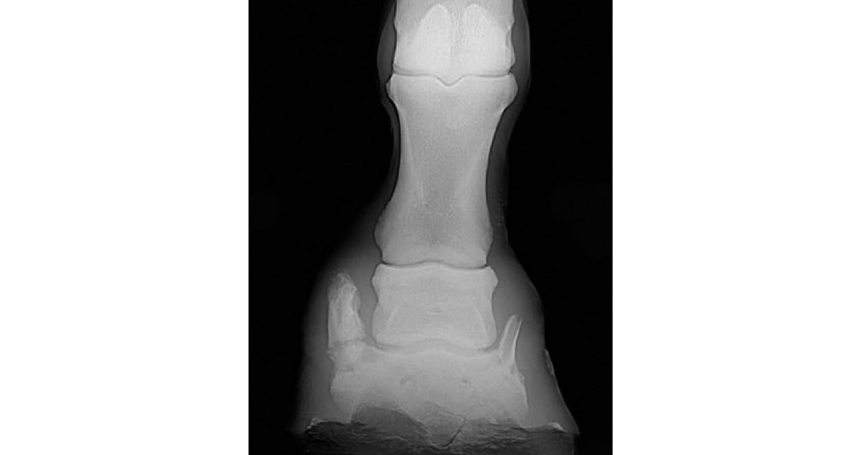
- Radiographic examination of the foot usually reveals the extent of the ossification of the cartilage or cartilages.
- Occasionally a sidebone may appear fractured; however, the radiolucent defect must be differentiated from the junction between a separate ossification center and that part of the cartilage that is ossifying from the palmar process of the distal phalanx.
- Asymmetrical swelling of pastern region, pain on palpation of the collateral cartilage, and improvement of the lameness with a uniaxial PD nerve block is suggestive of a problem in this region.
- Scintigraphy and/or MRI
Treatment
- If sidebone is suspected as the cause of lameness, conservative treatment with rest, topical 1% diclofenac sodium cream (Surpass®), and oral administration of NSAIDs is recommended initially.
- Any contributing foot problems such as foot imbalances should be addressed.
- Surgical removal of suspected fractured sidebones is not recommended. If lameness persists and sidebone is considered the cause of the lameness, a PD neurectomy can be performed but is usually unnecessary.
- Horses with sidebone and a secondary distal phalanx fracture are treated with corrective shoeing
Prognosis
The prognosis of ossification of the collateral cartilages of the distal phalanx is difficult to predict because this condition is thought to rarely cause lameness.