TABLE OF CONTENTS
Occipital Bone of Animals
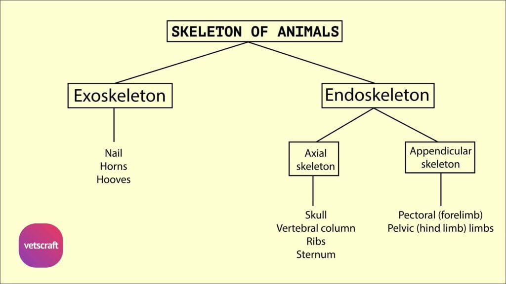
The occipital bone forms the caudal part of the skull in domestic animals and plays a crucial role in protecting the brain and forming joints with the vertebral column. It consists of several parts that vary among species like ox, horse, dog, and fowl.
Occipital Bone of Ox
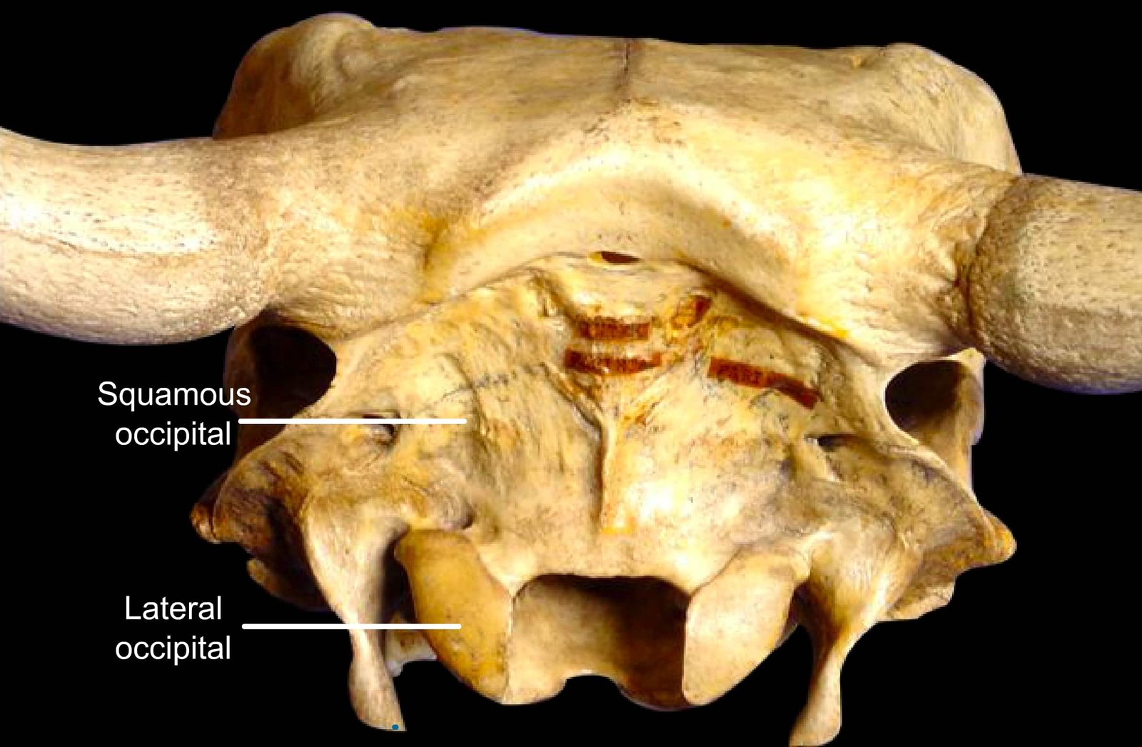
The occipital bone of the ox is a single bone situated on the lower part of the posterior surface of the skull. It consists of the lateral (exoccipital), squamous (supraoccipital), and basilar (basioccipital) parts.
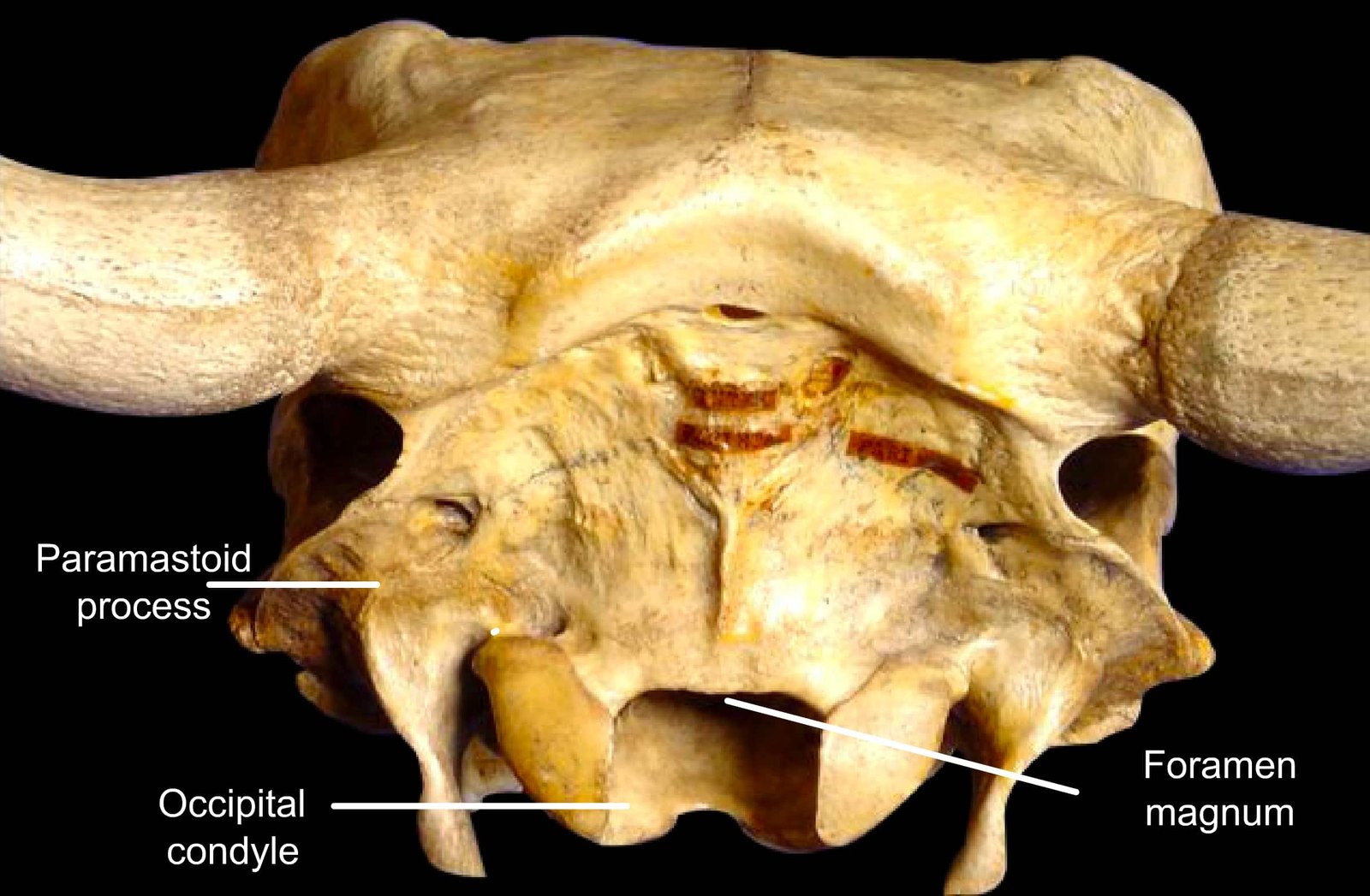
Lateral Part
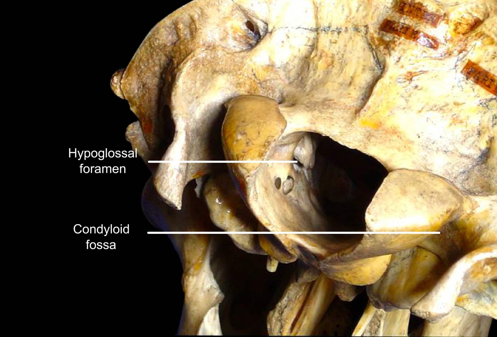
Each lateral part consists of a condyle and a paramastoid process. The condyles articulate with the atlas. Lateral to the condyle is the paramastoid process, which serves as a site for muscular attachment.
The paramastoid process projects downward and backward and is curved medially. Between the root of the paramastoid process and the condyle is the condyloid fossa, in which a large foramen—the hypoglossal foramen—is present for the XII cranial nerve. Above this is another foramen (often double) that conducts a vein from the condyloid canal.
The condyloid canal passes upward from a foramen on the medial side of the condyle and opens into the temporal canal.
The canal lodges a vein that connects the transverse sinus of the dura mater to the basilar venous plexus.
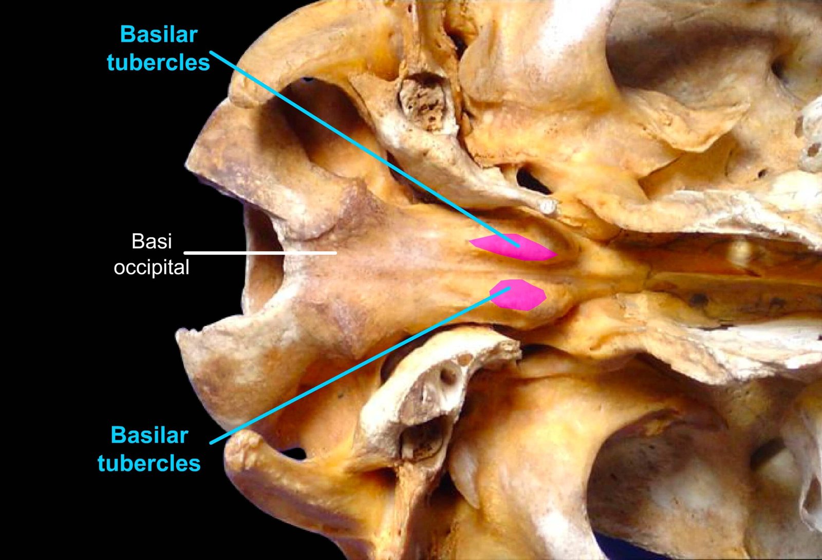
Basilar Part
The basilar part is a wide, thick bar of bone that extends forward from the ventral margin of the foramen magnum.
Its ventral surface is rounded, and its dorsal surface lodges the pons and medulla oblongata.
The anterior end is fused to the body of the post-sphenoid. At its junction with the post-sphenoid, it presents two external tubercles (basilar tubercles), which serve for muscular attachment.
The lateral borders form the medial margins of the foramen lacerum, which allows the passage of the IX, X, and XI cranial nerves.
The lateral and basilar parts enclose the large foramen magnum, where the cranial cavity and the vertebral canal join.
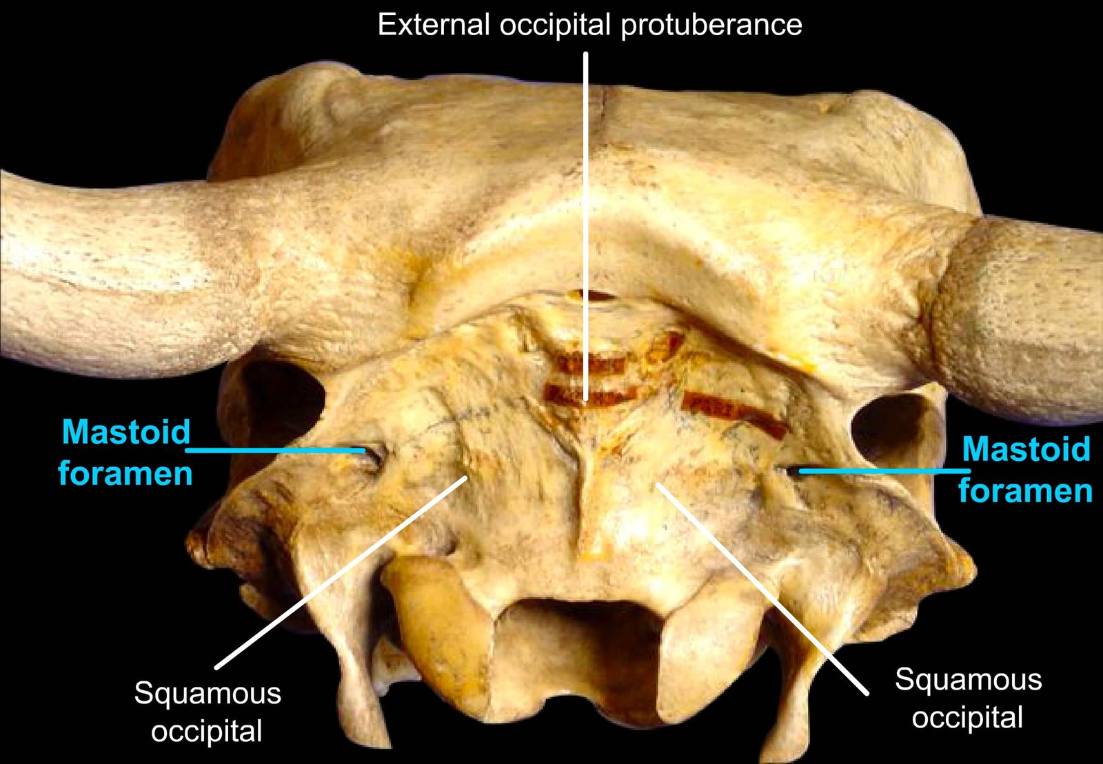
Squamous Part
The squamous part is a quadrilateral plate of bone lying between the lateral parts below, the squamous temporal laterally, and the parietal and interparietal bones, with which it fuses before birth.
Externally, it presents a central external occipital protuberance near its junction with the interparietal bone, which serves as the attachment for the funicular part of the ligamentum nuchae.
The mastoid foramen is situated on each side at the junction of the occipital and squamous temporal bones. It communicates with the temporal and condyloid canals at their junction.
The cerebral surface of the squamous part presents a shallow median fossa for the vermis of the cerebellum, and above this is a small eminence—the internal occipital protuberance.
A groove on either side (lodging the transverse sinus in life) leads into the temporal canal.
In the adult, it is excavated to form part of the frontal sinus.
Comparative Anatomy of Occipital Bone
The occipital bone exhibits notable anatomical differences among domestic animals. These variations reflect species-specific adaptations in skull structure, posture, and neurological function.
Horse
- The posterior surface of the skull is formed entirely by the occipital bone in the horse.
- The condyles in the lateral parts are obliquely placed.
- No other foramina besides the hypoglossal foramen are present in the condyloid fossa.
- The paramastoid process is longer, narrower, and less curved.
- The basilar part is longer and narrower, and the basilar tubercles are smaller.
- The foramen lacerum is wider and appears as a large triangular gap in the dry skull, but in life, it is covered for the most part by fibrous tissue (refer to the sphenoid for the foramina formed here).
- The squamous part forms the superoposterior part of the skull and is crossed externally by a prominent transverse and horizontal ridge called the nuchal crest.
- Its cranial surface shows a deep central depression and two shallower lateral depressions for the cerebellum.
Dog
- The occipital bone of the dog is situated as in the horse.
- The nuchal crest is prominent, angular, and directed backward.
- There is only one foramen in the condyloid fossa.
- The mastoid foramen and condyloid canal are as in the ox.
- The paramastoid processes are very short.
- The basilar part is wide and joins the bulla tympanica on either side.
- The basilar tubercles are located at the junction with the bulla.