Maxilla Bone of Domestic Animals: Ox, Horse, Dog and Fowl Anatomy
The maxilla bone is the principal bone of the upper jaw in domestic animals. It forms part of the face, nasal cavity, orbit, and hard palate, and carries the upper cheek teeth. The anatomy of the maxilla varies among species according to feeding habits and skull structure. This article describes the comparative anatomy of the maxilla bone in ox, horse, dog, and fowl.
Maxilla Bone of Ox
The maxilla bone of the ox is the largest bone of the upper jaw and carries the upper cheek teeth. It is situated on the lateral aspect of the face. It is composed of a body and two processes—the palatine and zygomatic processes.
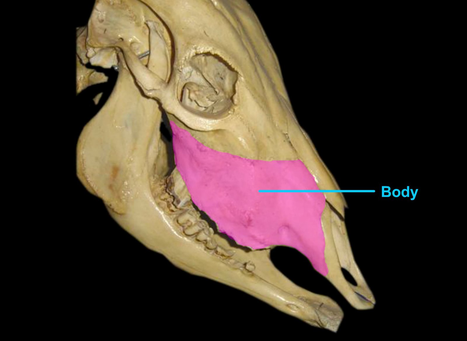
The external surface of the body is convex and presents, at the level of the first cheek tooth, the infraorbital foramen, which is the external opening of the infraorbital canal for the infraorbital artery and maxillary nerve.
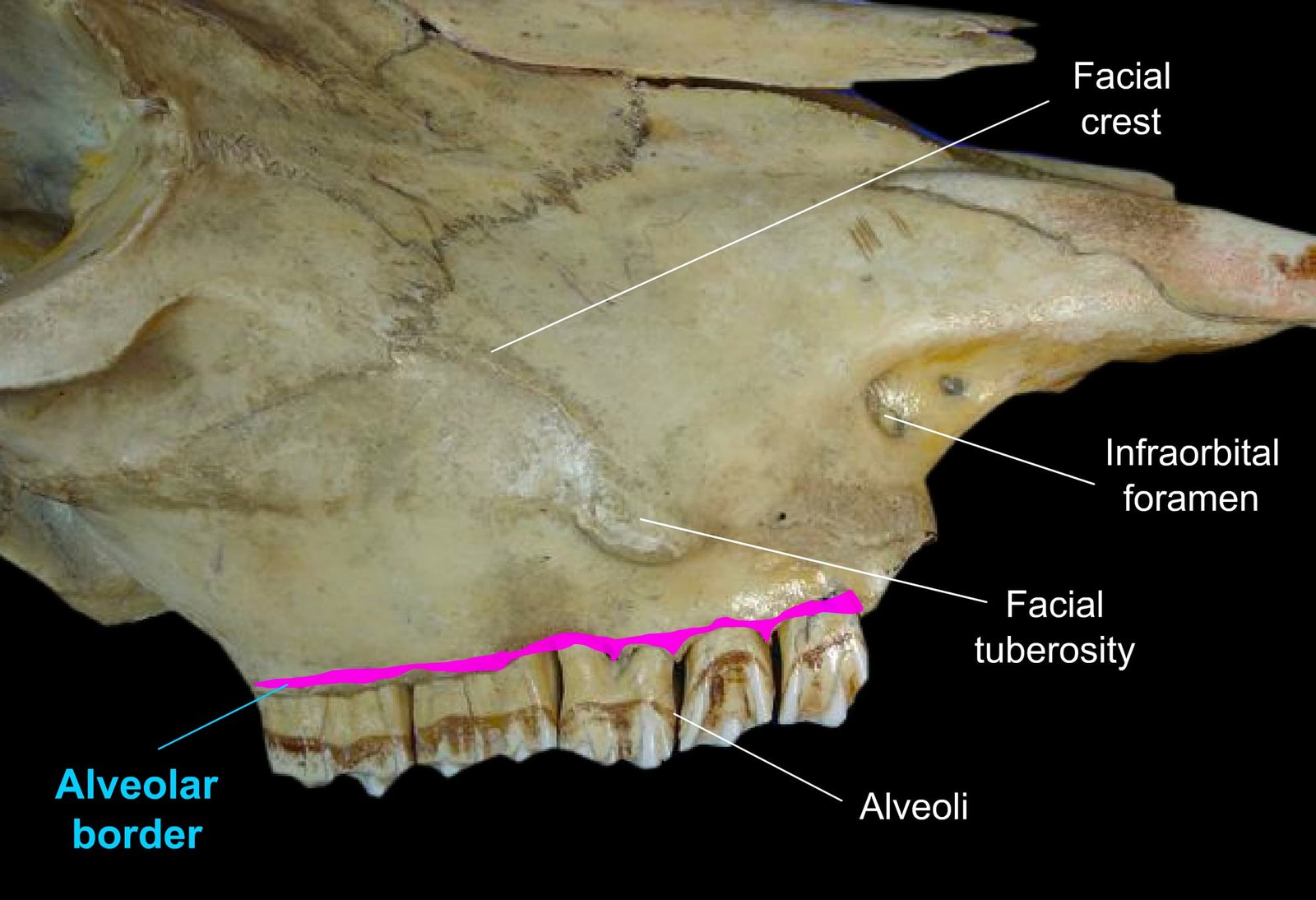
At the level of the third cheek tooth is the facial tuberosity, from which the facial crest extends.
The internal surface forms the lateral wall of the nasal cavity and presents, about the middle, the ventral turbinate crest for the ventral turbinate bone. Immediately below this is the lacrimal groove for the membranous nasolacrimal duct.
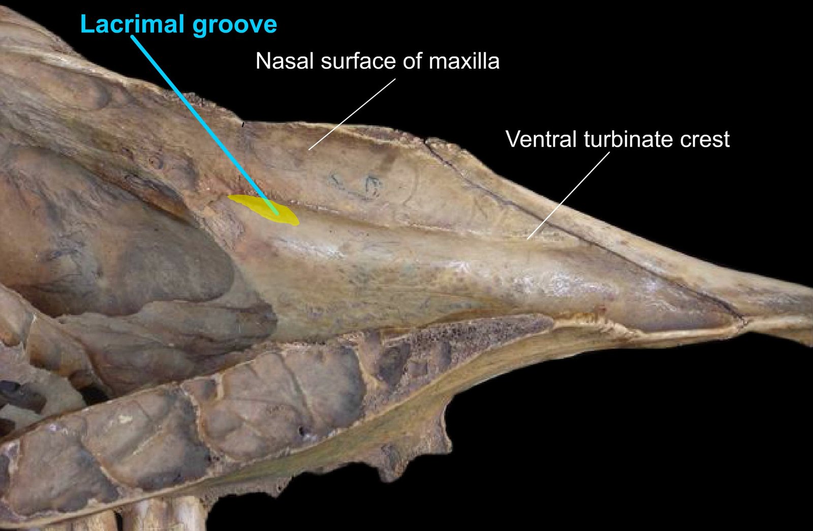
The dorsal border is irregular and scaly and articulates with the nasal process of the premaxilla, as well as the nasal, lacrimal, and malar bones. The ventral or alveolar border shows six alveoli posteriorly and is free anteriorly.
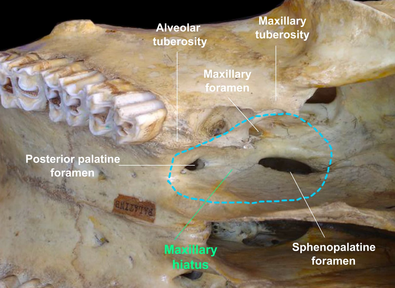
The anterior extremity is pointed and joins the premaxilla. Behind the last alveolus is a rough area called the alveolar tuberosity.
The maxillary tuberosity forms the posterior extremity of the bone and contains an extension of the maxillary sinus.
Medial to this is the maxillary hiatus, which presents the sphenopalatine, maxillary, and posterior palatine foramina for the nerves and vessels of the same names.
The zygomatic process is a small projection from the posterior extremity and is overlapped by the malar bone.
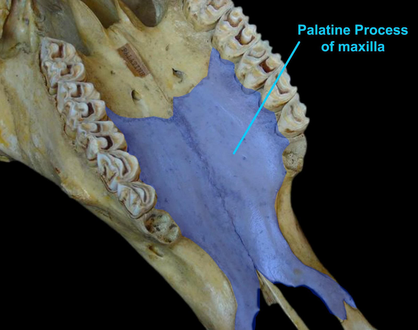
The palatine process forms a large part of the bony palate. Its oral surface shows the palatine groove for the greater palatine artery and nerve. It is excavated to form part of the palatine sinus.
Maxilla Bone of Horse
In the maxilla bone of the horse, the facial crest is prominent. The infraorbital foramen is located at the level of the third cheek tooth, and a small canal extends from it into the premaxilla.
The palatine canal is formed between the medial surface of the body of the maxilla and the vertical part of the palatine bone.
The medial borders of the palatine processes bear nasal crests on their nasal aspect, and these form a groove for the vomer. At the junction of the maxilla and premaxilla is an alveolus for the canine tooth.
Maxilla Bone of Dog
In the maxilla bone of the dog, the facial crest is absent. The infraorbital foramen is positioned similarly to that in the horse.
The frontal process fits into a deep notch between the nasal and orbital parts of the frontal bone. The maxillary and alveolar tuberosities are absent, but behind the last alveolus is a pointed pterygoid process.
There is a large alveolus for the canine tooth at the junction with the premaxilla.
Maxilla Bone of Fowl
In the maxilla bone of the fowl, the bones are slender and rod-like, placed on the sides and partly on the floor of the nasal cavity.
The two palatine processes do not meet but join the palatine and vomer posteriorly.
The maxilla joins the premaxilla and nasal bone anteriorly, while posteriorly it is continuous with the malar bone.