TABLE OF CONTENTS
Intestinal Obstruction in Animals
Intestinal obstruction in animals is an acute clinical emergency and can be caused by intestinal accidents like volvulus, intussusception, strangulation, or intra- or extra-luminal blockage. It is clinically characterised by signs of acute abdominal pain and the absence of defecation, followed by shock.
The common causes of intestinal obstruction in animals are volvulus, intussusception, strangulation, and intraluminal blockade. Other causes, like extra luminal masses causing pressure on the intestinal wall, may lead to compression of the lumen and blockage of the intestine.
Etiology
Intestinal accidents that cause intestinal obstruction are:
- Volvulus: twisting of the intestine on its own axis
- Tortion: twisting of the intestine along with its mesenteric axis. Usually, torsion is more common in blind sacculated organs, e.g., caecum, colon.
- Strangulation: constriction of a loop of intestine through a hernial ring. Eg: ventral hernia
Extra-luminal mesenteric lymphadenopathy, hydatidosis, or cysticercosis can cause luminal compression and distruction.
Intraluminal blockages are due to foreign bodies and are classified as:
- Phytobezoars: originating from plants
- Trichobezoars: originating from animal hair balls.
Pathophysiology
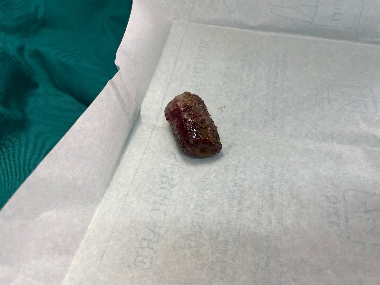
The enterolith is an obstructing mass in the intestine. The effect of intestinal obstruction differs from species to species due to the ability to sustain associated pain and toxaemia.
For example, acute intestinal obstruction of the small intestine can kill a horse in 24 hours. But similar situations do not result in the death of cattle for 6-7 days. A possible explanation for the above situation is the succeptibility of spp. to pain and the presence or absence of bacteria in the loop of the intestine. Once there is obstruction, further flow of ingesta is inhibited, and at the same time,intestinal secretion pours fluid into the lumen, causing acute distention of the lumen. The unfavourable organism colonises, releases endotoxins, and absorbs toxic metabolites into the circulation, resulting in profound shock, which has a direct effect on the cardiovascular and respiratory systems.
Overdistention causes stretching of nerve endings to cause severe abdominal pain, which is also an additional factor in the onset of acute shock. As the condition advances, dehydration leads to acid-base disturbance, resulting in peripheral circulatory failure.
Biochemical Changes due to obstruction of intestine
Loss of bicarbonate, hypochloremia, hypokalemia, hyponatremia, and, in later stages, hyperkalemia and elevated BUN take place in intestinal obstruction in animals.
Clinical Signs
Cattle kicking at the belly,uneasy treading of hind legs, and intermittent lordosis type appearance with bellowing. The animal may show frequent situps. Pain is spasmodic and occurs at regular intervals, and in some animals where pain is severe, it is indicated by rolling on the ground. Usually, there is accompanying anorexia. no defecation if faeces are present, palleted hard and pass coated with mucous and blood. Animal makes severe straining course in order to defecation. RR and HR are also elevated with varying degrees of dehydration. Per rectal examination reveals empty rectum and inserted hand is coated with mucous and blood.
As the condition worsens , animals are recumbent, showing signs of shock, congestion of the cyanotic mucous membrane,cold extremities, intermittent grunts, and low papillary reflex.
If the case is not treated in time, the majority of animals will die within 24–36 hours after the onset of shock.
In horses, the signs of acute intestinal obstruction are always clear and almost resemble those of equine colic, but the episode is much shorter, and signs of toxaemia and shock appear much earlier.
High PR of more than 80/min, RR of 60 to 80/min, HR of 100–120/min, accompanied by other signs such as sweating, panting type of respiration, and severe abdominal pain. Untreated cases enter profound shock within 12–24 hours. The medical treatment of the case at this stage is almost ineffective, indicating early surgical intervention.
Diagnosis
The diagnosis of intestinal obstruction is made by the following:
- Clinical signs of abdominal pain and an empty rectum by per rectal examination.
- Lab investigations reveal PCV > 55–55%, indicating hemoconcentration.
- Biochemistry reveals a decrease in bicarbonate, sodium, and chloride. Potassium initially decreased, but later hyperkalemia.
- Leucopenia usually indicates an infarction of the intestine or diffused peritonitis.
- Leucocytosis indicates leaking of contents in the peritoneum and cause peritonitis.
- Increased BUN indicates decreased kidney function due to ongoing toxaemia and hemoconcentration.
- Diagnostic imaging tools (ultrasonography) can be used to see an intestinal foreign body.
Differential Diagnosis
An intestinal obstruction case should be differentiated from the following diseases:
- Gastric dilatation in horses is not accompanied by the complete absence of faeces or the presence of blood and mucous stained faecal pallets. Nasogastric tube passing will specify the stomach contents.
- Peritonitis
- Renal colic or uroliths are similar, but oliguria or anuria is the most prominent sign rather than absence of defecation.
Treatment
Surgical removal of the obstruction is the only treatment for this. Supportive and symptomatic treatment can be given to manage and relieve pain. (See a small clip of surgery for the removal of intestinal obstruction from the intestine of a dog.)


