TABLE OF CONTENTS
Infectious Diseases Causing Infertility
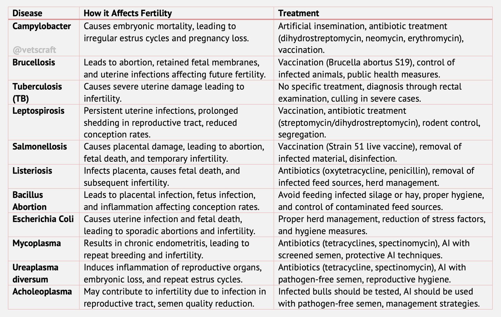
There is various infectious diseases causing infertility in animals like Campylobacteriosis, Brucellosis, Tuberculosis, Leptospirosis, Salmonellosis, Listeriosis, etc.
Specific infectious diseases causing infertility are:
- Campylobacteriosis
- Brucellosis
- Tuberculosis (TB)
- Leptospirosis
- Salmonellosis
- Listeriosis
- Bacillus Abortion
- Escherichia Coli
- Ureaplasma diversum
- Acholeoplasma
Campylobacteriosis
Campylobacteriosis also known as Vibriosis. It is caused by organism Campylobacter fetus. The bull carries the infection transmitting the infection at service to the female.
- Organism is confined to the glans penis, prepuce and distal urethra, with no lesions.
- Embryonic deaths
- irregular return to estrus.
The majority of the abortions due to C. fetus occur between the fourth and seventh months of gestation.
- There is necrosis, with yellowish brown discoloration of the fetal cotyledons and leather-like thickening or edema of the inter cotyledonary spaces.
Diagnosis
- History.
- semen of the newly introduced bull should first be eliminated.
- Diagnostic tests used to diagnose C. fetus infection include.
- Identification of the organism in preputial washings.
- Direct smears, culture and fluorescent antibody tests.
- Serological tests.
- Vaginal mucus agglutination.
Treatment and Control
- Infected bulls is replaced by artificial insemination.
- Removal of bulls from the herd.
- Regular testing of AI bulls.
- Dihydrostreptomycin at a dose rate of 22 mg/kg subcutaneously,
- A combination of neomycin and erythromycin,
- Vaccination should preferably be carried out 30-90 days before breeding.
- Vaccination has also been used to cure infected bulls. Two doses of vaccine at a month‘s interval, together with annual vaccination programmes, greatly reduces the incidence of genital vibriosis.
Brucellosis
Brucellosis is caused by brucella abortus in cows. Brucella melitensis in sheep and goats, can also be transmitted to cattle.
Epidemiology
Infection can be through:
- ingestion of B. abortus from contaminated pasture, food or water.
- licking an aborted fetus, infected afterbirth or genital exudates from a recently aborted or recently calved cow.
- teat by infected milk of another cow.
- vagina by infected semen.
The organism colonises the udder and supramammary lymph nodes of non-pregnant animals and infected cows often shed the organism in the milk, thereby endangering public health.
In pregnant animals, production of erythritol within the placenta allows rapid multiplication of the bacteria, leading to:
- endometritis, infection of cotyledons and placentitis
- abortions within 48 -72 hours after death of fetus, by which time a degree of autolysis has occurred.
- retention of fetal membranes.
Outside the animal body B. abortus may live for months in aborted fetuses or fetal membranes, but when exposed to drying and sunshine it is soon killed.
Calves that derive milk-borne infection throw off infection from the lymph glands of the gastrointestinal tract in 50-80 days.
Clinical Signs
- Results in abortion in the second half of pregnancy, together with metritis and retained fetal membranes.
- In bulls, it causes orchitis, epididymitis, seminal vesiculitis or infection of the ampullae.
- Infected cows usually abort once and seldom more than twice,
- Retained fetal membranes, delayed involution of the uterus leading to puerperal metritis.
Diagnosis
- Identification of organisms in stained smears.
- Fluorescent antibody technique for direct identification of the organism.
- Cultures from fresh afterbirth, or uterine exudates. Technique is time consuming and expensive.
- A colony blot ELISA using monoclonal antibodies provides a rapid, inexpensive and reliable method of identifying B. abortus.
- The Rose bengal plate test.
- A Serum Agglutination Test (SAT).
- The Compliment Fixation Test.
- The Milk Ring Test (MRT),
- The vaginal mucus agglutination test can be used on samples from individual cows but is not very reliable.
Control
- Brucellosis is not only a cause of abortion in cattle, but it also causes a serious disease, undulant fever, in man. Hence, control of the disease has to be directed at both its animal health and its public health aspects.
- By calfhood vaccination, using the B. abortus S19 live antigen. Vaccination
- Vaccinations are being done with Strain 19 a smooth variant of a strain of B. abortus, of reduced virulence but of high antigenic quality.
- Calves have to be vaccinated between 2 and 10 months of age.
- Vaccination of calves causes a febrile reaction and rapid sero conversion, with titres declining over the next 12 months in 90% of animals.
Tuberculosis (TB)
Uterine tuberculosis is of three clinical types- peritoneal, glandular and epithelial.
Diagnosis by rectal examination and the detection of thickened, tortuous tubes is diagnostic. In advanced cases, diffuse or nodular enlargement of the uterus will be readily detected.
Leptospirosis
Leptospirosis is caused by spirochaetes of the species Leptospira interrogans. It causes a zoonotic disease in man.
Mode of Transmission
- Infection can enter via skin abrasions or through the mucous membranes of the eye, mouth or nose.
- Transmitted in semen after natural service or AI.
- Organisms get localized in tissues that are inaccessible to antibodies, notably the kidney tubules, cotyledons and fetus.
Clinical Signs
- Leptospires can be present in puerperal discharges for up to 8 days and can persist in the pregnant and non-pregnant uterus for up to 142 and 97 days after infection, respectively.
- characterized by temperatures of 40°C or more, together with haemoglobinuria, icterus and anorexia is seen.
- Deaths may occur, especially in calves, and there may be abortions.
- In some herds, abortions have occurred after a ‗leptospiral mastitis‘ or agalactia has been observed during the previous 3 months. There is a precipitous fall in milk yield, especially in cows that are in early lactation.
- Milk from all four quarters is thick and colostrum like with clots, and is frequently blood-tinged known as “Gargetty milk”.
Diagnosis
There are no lesions that are specific for leptospirosis; thus diagnosis of leptospirosis as a cause of abortion is based almost entirely upon
- demonstrating specific antibodies in fetal sera
- by demonstrating leptospires in fetal organs, particularly lungs, kidneys and adrenal glands
- by culture or immunofluorescence.
The MAT is used extensively in the diagnosis of leptospirosis, using serum from animals that have aborted or are suspected of being infected.
Treatment and Control
- strict segregation of cattle from pigs,
- rodent control and the draining or fencing off of contaminated water sources.
- Sheep not to graze them together.
- There are two methods of specific treatment and control: the use of a vaccine or parenteral streptomycin/dihydrostreptomycin, or a combination of both.
- The antibiotic should be used at a does rate of 25 mg/kg by intramuscular injection with no greater a volume than 20 ml at any one site.
- In closed herds, vaccination of all members of the herd should be done annually. In open herds, the frequency should be increased to 6-monthly intervals.
Salmonellosis
Salmonellosis is caused by Salmonella typhimurium.
Pathogenesis
- S. Dublin, the organism rapidly spreads to the liver, spleen, lungs and adjacent lymph nodes of the dam;
- Pyrexia. Six to eight days later it spreads to the placentomes,
- The placentome is damaged, probably by endotoxin, causing necrosis, placental failure, fetal death and abortion.
Clinical Signs
- Marked pyrexia severe diarrhoea and dysentery, which may be associated with abortion. More frequently, salmonella abortions occur in late pregnancy although malaise, pyrexia and inappetance have also been recorded.
- RFM is a common sequel, although there is no adverse effect upon fertility.
Diagnosis
- A definite diagnosis depends upon the isolation of the organism from fetal tissues and membranes, uterine discharges or vaginal mucus. Serological tests can be used, especially the SAT, although agglutinins fall to low titres fairly soon after the event.
Control
- Cows that have aborted only excrete the organism for a very short period of time, unlike the continuous or intermittent excretors that occur following enteric infection.
- fetuses and fetal membranes together with contaminated bedding should be disposed of safely. Adequate cleansing and disinfection of premises should be performed.
- S. Dublin can be controlled by vaccination with the Strain 51 live vaccine.
- Killed vaccines and bacterins have also been used, largely against S. typhimurium.
Listeriosis
Listeriosis is caused by Listeria monocytogenes. It is isolated from bovine abortuses, and is also a cause of abortion in sheep and goats.
Pathogenesis
- The organism gains entry by ingestion or by penetration of mucous membranes of the respiratory system or conjunctiva, as well as the central nervous system.
- L. monocytogenes has a predilection for the placenta, causing placentitis, and affects the fetus to cause abortion.
Transmission
- L. monocytogenes being present in the soil, sewage effluent, bedding and foodstuffs;
- There is good evidence that there is an association between listeriosis and the feeding of poor-quality silage of higher-than-normal pH.
- Cross-infection between sheep and cattle is possible.
Clinical Signs
- Usually abortions are sporadic, occurring towards the end of gestation. However, there are rare reports of serious outbreaks, or abortion storms in some herds.
- Pyrexia before, at the time of or after abortions have occurred. The aborted fetus frequently has characteristic multiple yellow or grey necrotic foci in the liver and cotyledons.
Diagnosis
- Identification of the organism in the abomasum and liver of the fetus, and in the placenta and vaginal discharges by a direct smear or by immunofluorescence.
- Culture of the organism is not easy, although a series of sub cultures following refrigeration has proved to be successful.
- Serological tests are not used in its diagnosis.
Treatment and Control
- The possibility of preventing further abortions occurring in a herd might be considered by using oxytetracycline or penicillin; however, this is rarely practicable. If silage is being fed this must be considered to be a potential source of infection and, if possible, withheld from pregnant cows. There is evidence that some individuals become symptomless carriers, excreting the organism in faeces and milk.
Bacillus Abortion
Bacillus abortion caused by Bacillus licheniformis.
Clinical Signs
- The placentitis due to B. licheniformis is similar to that following mycotic infection.
- The allantochorion is dry, leathery and yellow or yellowish brown in colour.
- There is often edema of the allantochorion, especially around the cotyledons, which appears almost as if there are vesicles present.
- The cotyledons are haemorrhagic and necrotic. The fetus may be infected and, if so, there will usually be evidence of a fibrous pleurisy, pericarditis and peritonitis.
- There are no systemic signs of disease in the cow.
Diagnosis
- This depends upon the appearance of the placenta and the culture of the Bacillus in placenta and vaginal swab.
Transmission and Pathogenesis
- Common source of infection is silage, especially when water, other foodstuffs and bedding are contaminated with silage effluent.
- Wet, spoilt hay can also be a source.
Control
- Infected silage or hay should not be fed.
Escherichia Coli
Sporadic abortions due to E. coli have been reported. It is suggested that, following stress, the organism reaches the fetus and placenta via haematogenous spread or ascending the genital tract.
Mycoplasma
- M. bovigenitalium is found in the vaginal mucus of normal and repeat breeder cows, which has led to speculation concerning its role as a pathogen.
- The organism may also cause granular vulvovaginitis.
- M. bovis causes mastitis in adult cattle and polyarthritis in calves. causing extensive lesions of the uterus, uterine tubes and even peritonitits.
Ureaplasma diversum
- Ureaplasma diversum is persists only briefly in the uterus and uterine tubes, but is most commonly found in the vagina and vestibule.
- Condition attributed to U. diversum infection is granular vulvovaginitis.
- Acute infection produces granules around the clitoral region and on the lateral walls of the vagina, which are accompanied by hyperemia of the vulva and a profuse, mucopurulent vaginal discharge.
- Large, purulent lesions may also be present, which resemble those of IPV.
- U. divrsum produce endometritis and salphingitis. These lesions have been associated with high levels of embryonic death and returns to estrus, which are accompanied by a mucopurulent vaginal discharge. Abortions may also occur.
- U. diversum can infect the penis and prepuce of the bull and has occasionally been isolated from all parts of the male tract. It is generally regarded as non-pathogenic in the male.
- Transmission of the infection is by the venereal route. Infected semen used in AI seems of particular importance, since its deposition into the uterus allows the development of chronic endometritis, rather than of acute vulvovaginitis.
Acholeoplasma
- Three species of Acholeplasma have been isolated from cattle. A. modicum, A. laidlawii and A. axanthum. Of these, a laidlawii has been isolated most often, largely from the bull.
- It is possible that Acholeplasma infection of cows may cause pathological changes in the genital tract, but the case is far from proven.
- It is often isolated from aborted calves, but as described above, may not be the cause organism. It probably causes no pathological lesions of the bull.
Transmission
- Spread of the organism from infected bulls and resultant infertility have also been demonstrated. M. bovigenitalium also inhabits many parts of the reproductive tract of the bull.
- When it infects the testes or epididymides, M. bovigenitalium may cause detrimental changes to semen quality, especially after cryopreservation.
- Isolation of the organism from the placenta or aborted fetus can be considered significant.
- M. bovis is found in bovine semen less often than M. bovigenitalium.
Diagnosis
- Most bovine mycoplasmas are easily recovered in conventional mycoplasma media,
- The development of ELISA and other diagnostic tests is likely in the near future.
Treatment and Control
- Natural service, if used, should be suspended and semen should be collected and cultured for the presence of mycoplasmas.
- Instead, animals should be inseminated with semen that is known to be free of contaminant organisms.
- Infected bulls should be rested for 3 months and treated systemically for 5 days with tetracyclines, together with sheath irrigation.
- A combination of lincomycin, spectinomycin, tylosin and gentamycin, spectinomycin, added to raw semen, and non-glycerolated whole milk or egg yolk-based extenders has been shown to control M. bovis, M. bovigenitalium and Ureaplasma spp.
- If artificial insemination is used, the standard Cassou pipette should be protected by a disposable polythene sheath to prevent vulval or vaginal contamination before it is introduced through the cervix.
- The uterus can be infused with a solution containing 1 g of tetracycline or spectinomycin 1 day after insemination, a treatment that has been shown to improve pregnancy rates.
- Transfer to pasture of affected animals should be considered. This may reduce spread by direct contagion.

