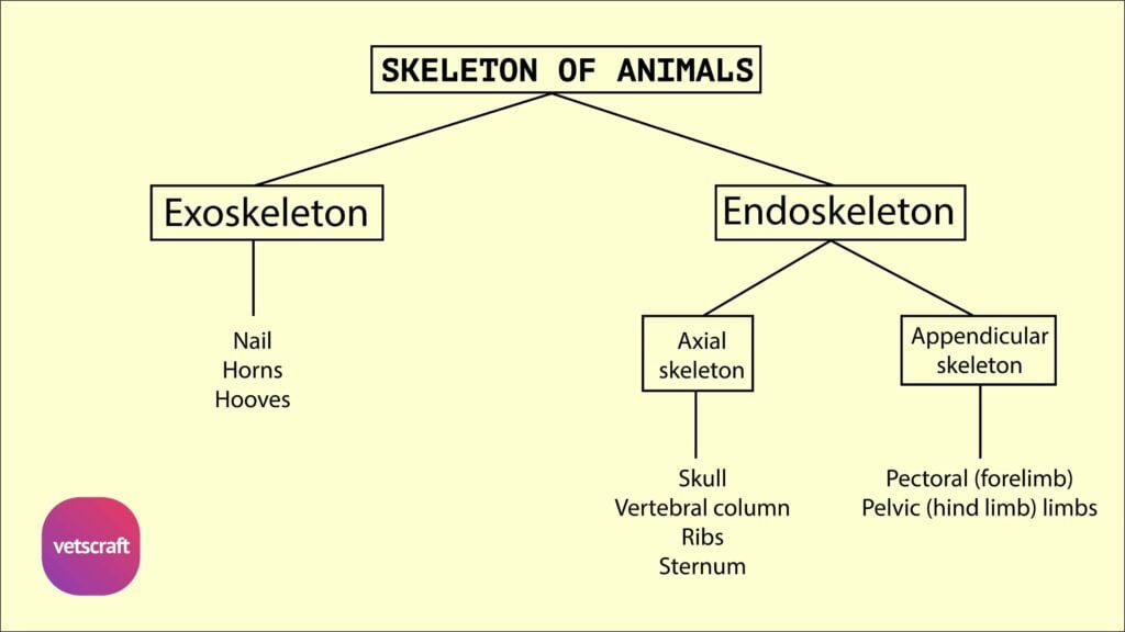
The Thyroid gland is surrounded by a connective tissue capsule, which gives off septa of varying thickness into the interior. These divide the organ into interconnected lobule consists of vesicles of varying sizes called thyroid follicles.
In young animals they are smaller than in adult. Between follicles there is fine connective tissue fibres, which contain many blood vessels and capillaries.
The follicles are completely enclosed and surrounded by fine reticular fibres. They are usually spherical or ovoid.
Cells lining these are cuboidal with large round nuclei. The cells may be flattened or squamous in distended follicles. The epithelium rests on a thin, delicate basement membrane.
The cavity of the follicle is filled with a semifluid or gel-like substance, the thyroid colloid. The colloid generally stains acidophilic but may be basophilic especially in activated glands. In young animals, between the follicles, small groups of cells may be seen. These are the primitive or embryonic cells, which may give rise to more number of follicles.
The secretion produced by the cells is stored in the vesicles as thyroglobulin.
The thyroglobulin is hydrolysed by enzymatic action of cells and the thyroxine is secreted into the capillaries at the base of the cells.