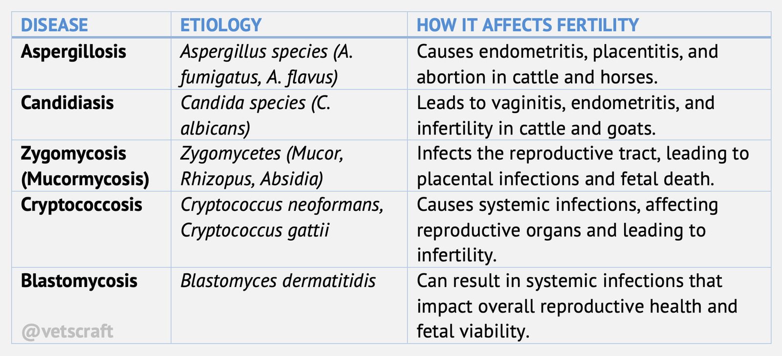
Fungal Diseases Causing Infertility
Fungal diseases causing infertility in animals by invasion of the placenta and fetus is a frequent and consistent cause of abortion In cattle.
Abortions are normally sporadic, although in some herds the incidence may be as high as 5-10%.The fungi that are most frequently isolated following abortion are Absidia sp., Rhizopus sp and Aspergillus sp.
Other fungi such as Mortiella wolfii and Petriellidium boydii have also been implicated. In the northern hemisphere, A fumigatus is the most common cause of abortion, while in the southern hemisphere, M. wolfii is the most important organism.
Transmission and Pathogenesis
Many of the species of fungi are ubiquitous in the air and environment in which cattle live; however, there is good evidence that mouldy hay and straw and other food such as silage and sugar beet pulp are important sources of infection.
Mycotic abortion is most prevalent in the winter months when cattle are housed.
It is generally agreed that there is haematogenous spread following entry into the vascular system from the respiratory or alimentary tracts.
There is some evidence that fungal-contaminated semen can cause uterine lesions although this route is unlikely to be important.
Clinical Signs
- Infection does not always cause abortion, since infected live calves can be born.
- abortions occurring between 4 and 9 months.
- The whole or part of the placenta usually appears discolored when shed, and is either grey, yellow or reddish-brown; the intercotyledonary areas of the allantochorion are thickened, wrinkled or leathery.
- placenta has been shed appear thickened and have a cup-like or coffee bean appearance. Characteristic fetal skin lesions include circumscribed, grayish-white thickened patches.
Diagnosis
The appearance of the placenta is fairly typical in fungal abortion, although some bacteria can produce similar lesions. The fetal skin lesions are almost pathognomonic.
Laboratory confirmation requires submission of placental tissue, Culture from fetal lungs and abomasum is more reliable but contamination can occur.
The reliable and traditional method of diagnosis is the identification of fungal cells in histological sections of the placenta.
Another technique is the potassium hydroxide ‗crush‘ mount of non-fixed tissue.
Conclusive diagnosis of mycotic placentitis can be made if:
- the characteristic lesions of placentitis are present in association with the presence of mycotic elements.
- the characteristic lesions of fetal dermatomycosis are present in association with the presence of mycotic elements.
- there is a fetal bronchopneumonia associated with mycotic elements.
- Serological tests are at present, unreliable and cannot be used for routine diagnosis.
Control
The feeding of mouldy forage or the use of mouldy bedding should be avoided.

