TABLE OF CONTENTS
Fibrotic and Ossifying myopathy in Horses
Fibrotic and Ossifying myopathy in Horses is a fibrosis with or without ossification of the muscle tissue in the crus that often results in adhesions between the semitendinosus, semimembranosus, gracilis, or biceps femoris muscles.
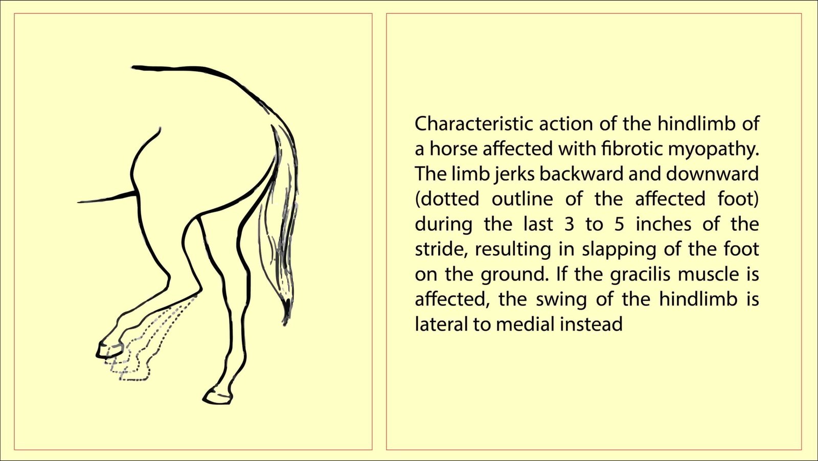
Fibrotic and Ossifying myopathy in Horses is significant because the fibrosis and adhesions limit the action of the semitendinosus muscle, causing an abnormal gait characterized by a slapping down of the foot at the end of the cranial phase of the stride. If the semitendinosus is more affected, the slap is cranial to caudal. If the gracilis is more affected, the slap is from lateral to medial.

The affected hindlimb approaches the ground more vertically than normal and strikes it toe-first, making more of a slap than the normal heel-toe manner of landing. Some have reported a semi-flexed stance at rest with the toe touching the ground. Rarely, the condition occurs in the forelimb.
A congenital form of fibrotic myopathy-type syndrome has been reported. Foals are born with an altered gait characteristic of fibrotic myopathy in the hindlimb. On palpation, a tightening of the semitendinosus muscle was appreciated but no firm thickening of the muscle typical of fibrotic myopathy was palpated.
Etiology
- The causes of fibrosis is always trauma
- Usually unilateral, rarely bilateral fibrosis was reported
Clinical signs
The signs are due to lack compliance of the affected muscle(s) and adhesions between the semitendinosus muscle and the semimembranosus muscle medially, and between the semitendinosus and the biceps femoris muscle laterally.
These adhesions partially inhibit the normal action of the muscles.
In the cranial phase of the stride, the foot of the affected hindlimb is suddenly pulled caudally 3 to 5 inches just before contacting the ground. The cranial phase of the stride is shortened, so consequently the caudal phase is lengthened.
Usually the lameness is most noticeable when the horse walks.
An area of firmness can often be palpated over the affected muscles on the caudal surface of the affected limb at the level of the stifle joint and immediately above it.
Occasionally, the lesion is deep and difficult to palpate or it may be in the medial gaskin affecting the gracilis muscle.
If the gracilis is affected, the slap motion of the foot is in a more lateral to medial direction.
Diagnosis
The diagnosis is based upon the altered gait and palpation of a hardened area on the caudal or caudomedial surface of the limb proximal to the level of the stifle joint. In making the diagnosis, stringhalt should also be considered
Treatment
Three approaches for surgical correction of fibrotic myopathy have been described; each must be adjusted if more than the semitendinosus muscle is involved-
- Complete removal of the affected fibrotic/calcified muscle tissue
- A semitendinosus tenotomy
- At the level of its insertion on the proximal medial tibia caudal to the saphenous vein. The procedure is much less invasive and is expected to be effective if the problem is unaffected by other muscles. To perform the semitendinosus tenotomy, the horse is positioned in lateral recumbency with the affected limb down. The tibial insertion of the semitendinosus tendon is palpated caudomedial to the proximal end of the tibia. An 8-cm incision is created over the tendon to expose it. The tendon is then isolated with a hemostat and transected. Hand-walking is started when the skin has healed, and normal exercise is allowed after 6 weeks.
- Focal semitendinosus myotomy
- Caudal thigh region where the myotomy procedure is performed to correct fibrotic myopathy. The skin incision is made vertically and the fibrotic muscle is transected with a bistoury horizontally.
Prognosis
The prognosis is good to guarded.