TABLE OF CONTENTS
Femur Bone of Domestic Animals: Ox, Horse, Sheep, Goat, Pig, Dog, Rabbit, and Fowl
The femur is the longest and strongest bone in the body, forming the skeleton of the thigh region in animals. It extends from the hip joint (acetabulum) to the stifle joint (knee), connecting the pelvic limb to the lower leg.
The femur consists of a shaft, a proximal extremity (with the head and trochanters), and a distal extremity (with the condyles and trochlea).

Femur Bone of Ox
The femur of the ox is the most massive of the long bones, extends obliquely downward and forward between the hip and stifle joints. It consists of a shaft and two extremities.

Shaft
The shaft possesses four surfaces. The anterior, medial, and lateral surfaces are continuous, convex from side to side, and are covered in life by the quadriceps femoris.
The posterior face is narrow in the middle, where it is rough for the adductor. Below this is an oblique vascular impression running downward and outward, marking the course of the femoral vessels.
The medial border of the posterior surface presents, in its upper third, the trochanter minor, which is for the quadratus femoris and ilio-psoas. Extending from this trochanter obliquely and joining the trochanter major is the trochanteric ridge, which forms the postero-lateral boundary of the trochanteric fossa. The ridge is for the gluteus medius, and the fossa is for the gemellus, obturator externus, and obturator internus.
The distal third of this border carries, above the medial condyle, the medial supracondyloid crest for the medial head of the gastrocnemius. The rest of this border, below the trochanter minor, is for the pectineus.
The lateral border presents, in its distal third, the supracondyloid fossa, which is bounded laterally by the lateral supracondyloid crest. The fossa is for the superficial flexor of the digit, and the crest is for the lateral head of the gastrocnemius.
Proximal Extremity
The proximal extremity is composed of the head and the trochanter major.
The head is medial and articulates with the acetabulum. The small non-articular sulcus, fovea capitis, in the middle of the head is for the round ligament of the hip joint.
The trochanter major, or greater trochanter, is massive and serves as the attachment for the gluteus medius. The lateral face is convex. Below its base are two rough tubercles—the upper one for the middle gluteus and the lower one for the deep gluteus.
Distal Extremity
The distal extremity is large and comprises the trochlea in front and two condyles behind. The trochlea articulates with the patella. The medial ridge of the trochlea is more prominent.
The condyles are separated by the intercondyloid fossa and articulate with the condyles of the tibia through the medium of the interarticular cartilages or menisci.
The medial condyle presents an eminence on its medial aspect for the medial ligament.
The lateral condyle presents two depressions on its lateral aspect: the upper one for the lateral ligament of the stifle and the lower one for the popliteus.
Between the lateral condyle and the lateral ridge of the trochlea is the extensor fossa for the complex muscle.
The intercondyloid fossa lodges the spine of the tibia. Its anterior part is for the posterior cruciate ligament. At its posterior part, close to the medial condyle, is a depression for the coronary ligament of the lateral meniscus, and close to the lateral condyle is another depression for the anterior cruciate ligament.
Comparative Anatomy of Femur Bone in Animals
Comparative anatomy of the femur bone in animals involves studying how the structure of the femur in various domestic species—such as the horse, sheep, goat, pig, dog, rabbit, and fowl—differs from or resembles that of the ox.
Sheep and Goat
- A distinct line separates the lateral and posterior surfaces of the femur in sheep and goats.
- The trochanter major is slightly higher than the head.
- The trochlear ridges are slightly oblique.
Horse
- The femur bone of the horse is more massive.
- The posterior face bears, in its proximal third, a rough eminence for the biceps femoris.
- The trochanter minor is in the form of a rough ridge.
- The lateral border bears the trochanter tertius in its proximal third for the superficial gluteus.
- The supracondyloid fossa and the lateral supracondyloid crest are better developed.
- The trochanteric ridge is vertical and extends from the proximal third to the great trochanter.
- The great trochanter is made up of a convexity, a summit, and a crest.
- The crest is below and lateral to the convexity.
Pig
- The shaft is wide and relatively massive in the femur of pigs.
- A ridge extends from the trochanter major to the lateral supracondyloid crest, and there is no supracondyloid fossa.
- The third trochanter is absent.
Dog
- The shaft of the femur in dogs is proportionately large and strongly curved, with the convexity forward.
- The supracondyloid fossa is absent.
- The trochanteric fossa is rounded and deep.
- The ridges of the trochlea are sagittal and equal.
- The intercondyloid fossa is wide.
- On the posterior aspect of the distal extremity, immediately above each condyle, is a small facet for a sesamoid—the fabella.
- The fabellae are two small, rounded sesamoid bones, located one on each condyle of the femur on the posterior aspect.
- They are developed in the tendons of origin of the gastrocnemius muscle.
Rabbit
- Immediately below the head is the trochanter minor, and lateral to it is a pair of projections, which form the greater trochanter and third trochanter in the femur of the rabbit.
- The distal extremity has medial and lateral condyles, separated by a median patellar groove.
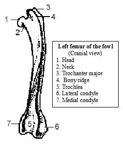
Fowl
- The head is prominent but smaller than the acetabulum, and the articular surface extends onto the trochanter and articulates with the acetabulum and the facet on its rim in the femur of the fowl.
- The lateral condyle presents, on its lateral aspect, a groove for the head of the fibula.