TABLE OF CONTENTS
Ethmoid Bone of Domestic Animals
The ethmoid bone in domestic animals such as ox, horse, dog, and fowl plays a vital role in separating the cranial and nasal cavities. It is a single, sieve-like bone composed of the cribriform plate, perpendicular plate, and paired lateral masses.
Ethmoid Bone of Ox
The ethmoid is a single bone situated in front of the pre-sphenoid and has a cribriform plate, a perpendicular part, and two lateral masses.
Cribriform Plate
The ethmoid is a sieve-like partition between the cranial and nasal cavities. Its cranial surface is divided by the ethmoidal crest (crista galli) into two halves.
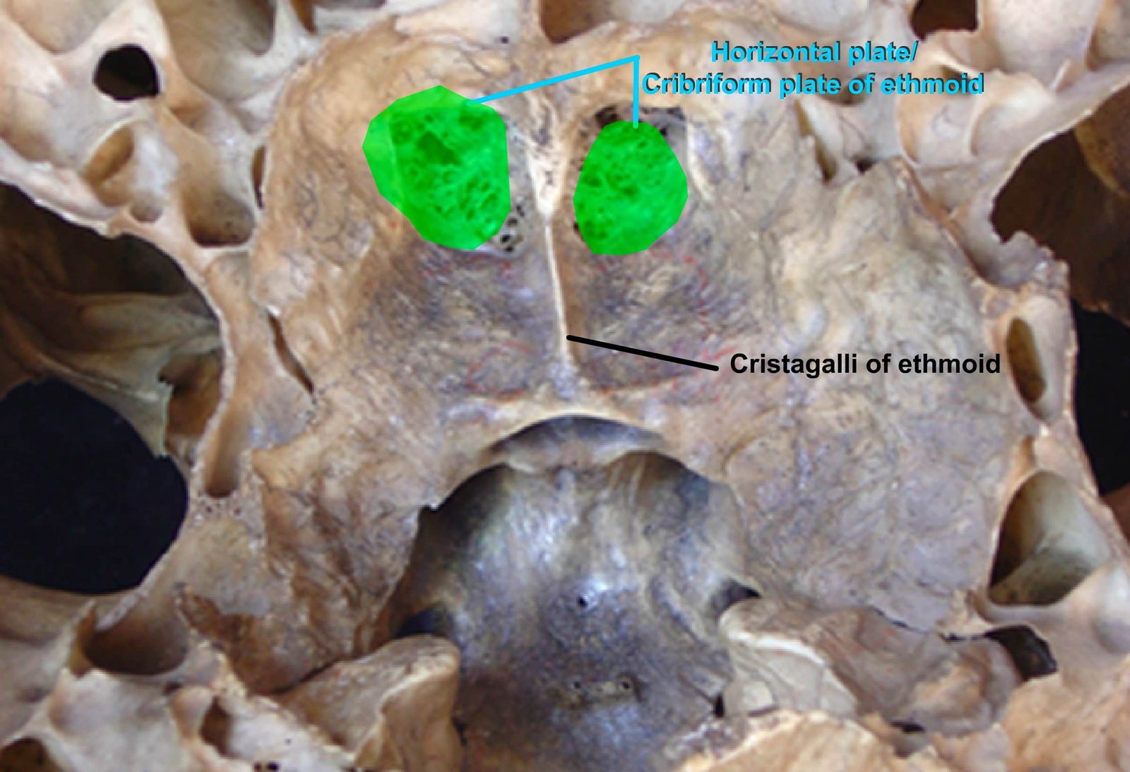
Each half forms the deep ethmoidal fossa for the olfactory bulb. The plate is perforated by numerous small foramina for the passage of the olfactory nerve filaments, and on either side is the ethmoidal foramen for the ethmoidal artery and nerve.
The convex nasal surface has the lateral masses attached to it.
Perpendicular Plate
The perpendicular plate forms the posterodorsal part of the septum nasi and is covered by a mucous membrane. Its dorsal border joins the junction of the frontals.
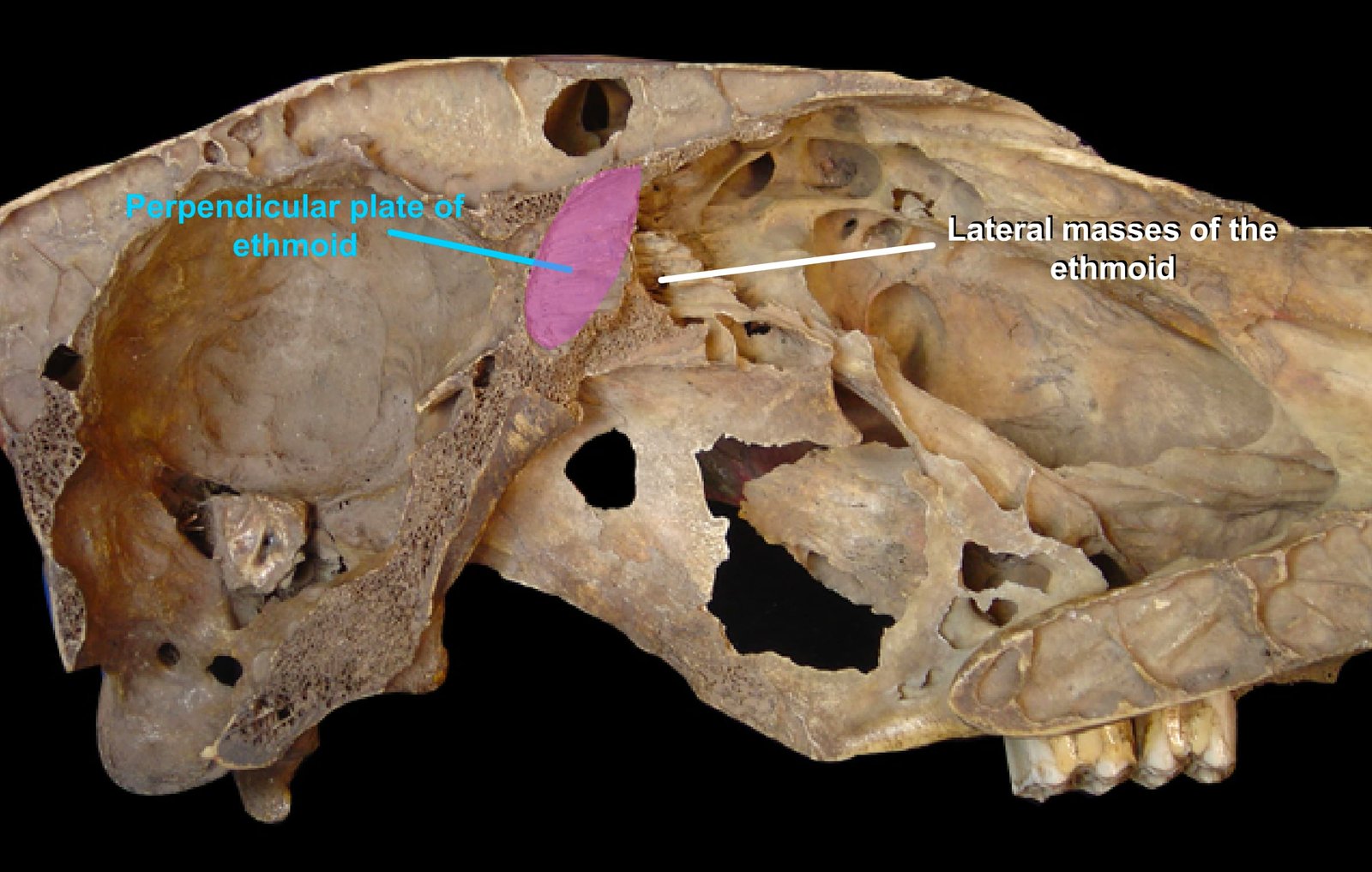
Its ventral border is received into the groove of the vomer, and its anterior border is continuous with the septal cartilage in life.
Lateral Mass
Each lateral mass forms the posterior part of the nasal cavity above and behind the posterior nares. The perpendicular plate separates the two masses.
Each has the shape of a cone, with the base attached to the nasal surface of the cribriform plate. Each is curved on its lateral face by a thin plate of bone, the lamina lateralis.
The ventral part of this lamina is seen in the pterygo-palatine fossa, forming the dorsal margin of the sphenopalatine foramen.
The mass consists of delicate, scroll-like plates of bone termed ethmo-turbinates. They are attached to the lamina lateralis and are separated by narrow intervals termed ethmoidal meatuses, which communicate with the nasal cavity.
In the fresh state, the ethmo-turbinates are covered with mucous membrane.
The largest ethmo-turbinate is so extensive that it projects between the dorsal and central turbinates and is often called the middle or third turbinate bone.
The mucous membrane covering the ethmo-turbinates is part of the olfactory mucous membrane.
Comparative Anatomy of Ethmoid Bone
The ethmoid bone varies significantly among domestic animals in both structure and development.
Horse
- The great ethmo-turbinate is less massive in the horse.
Dog
- The ethmoid bone is highly developed in dogs.
- The cribriform plate is extensive, and the olfactory fossae are very deep.
- The ethmoidal crest is poorly developed and often incomplete.
- The perpendicular plate is long.
- The lateral masses are greatly developed and project into the frontal sinus.
- The lamina lateralis is extensive and forms the medial wall of the maxillary sinus.
- Its ventral border joins the palatine process of the maxilla and the horizontal part of the palatine.
- A shelf-like plate extends inward from its lower part, connects with the similarly incurved part of the palatine bone, and meets the outcurving plates of the vomer to form the lamina transversalis, which divides the olfactory fundus of the nasal cavity from the lower nasopharyngeal meatus.
Fowl
- The lateral masses and the horizontal part are absent in fowl.
- The perpendicular plate forms the interorbital septum with the sphenoid