TABLE OF CONTENTS
Predominant Sympathetic or Parasympathetic Tone in various structures
| Site | Predominant Tone |
| Arterioles | Sympathetic (Adrenergic) |
| Veins | Sympathetic (Adrenergic) |
| Heart: atrium SA node Ventricle | Parasympathetic (Cholinergic) Parasympathetic (Cholinergic) Sympathetic (Adrenergic) |
| Iris | Parasympathetic (Cholinergic) |
| Ciliary Muscle | Parasympathetic (Cholinergic) |
| G.I. Tract | Parasympathetic (Cholinergic) |
| Urinary Bladder | Parasympathetic (Cholinergic) |
| Salivary Gland | Parasympathetic (Cholinergic) |
| Sweat Glands | Sympathetic (Adrenergic) |
In organs receiving both sympathetic and parasympathetic innervations the effects of the two divisions are usually opposed or antagonistic. Example: Heart, bladder, bronchi, GI tract (even in these organs one division is usually predominant)
In organs receiving dual innervations with the influences of the two divisions in the same direction and effects are complementary. Example: salivary glands ( salivary secretion is viscous and scanty with sympathetic tone and profuse and watery with parasympathetic tone)
In organs receiving only single innervations from one or the other division of ANS, the neuronal control level of function is by increasing or decreasing activity.
There are no parasympathetic innervations to systemic blood vessels, erector pili, sweat glands, adrenal gland and plain muscle of the upper eyelid and nictitating membrane
In some organs, the control of functions is regulated by, the opposing branches of the ANS. But each branch exerts its activity on different cells. Example: pupil.
The dilator and constrictor muscles of the pupil are kept in a state of tonic contraction by the sympathetic and parasympathetic nerves respectively.
In dim light, the pupil dilates because of reduced activity in the parasympathetic supply.
In a frightened animal the pupil dilates even in bright light, due to increased activity in the sympathetic nerve fibers.
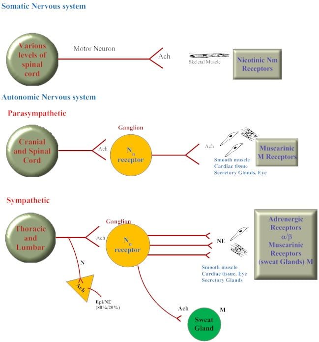
Distribution of cholinergic and adrenergic neurons
Cholinergic receptors
- All motor nerves to skeletal muscle
- All preganglionic autonomic nerves (including those to adrenal medulla)
- All postganglionic parasympathetic nerves
Adrenergic receptors
- Most postganglionic sympathetic nerves
- Innervations to cardiovascular system
