Development of Teeth of animals
Development of Teeth of animals take place by the both ectoderm and mesoderm.
A tooth is a highly modified connective tissue papilla that has undergone a peculiar ossification into dentine and caged by hard enamel elaborated from the epidermis. In addition the cement a bony deposit encrusts the base. The enamel is from ectoderm arid the dentine pulp and cement are from mesoderm.

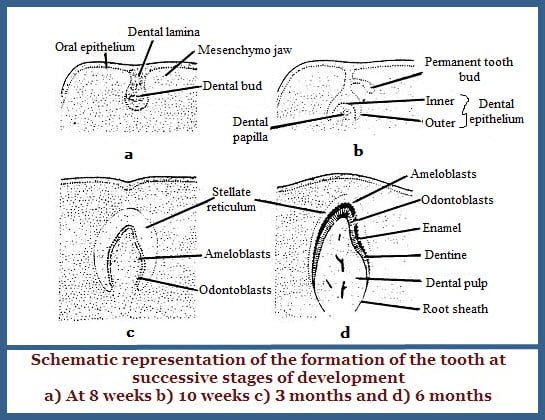
The earliest indication of the development of teeth is the appearance of a dental groove on the surface of the gum about the seventh week of the embryo. From this groove a lamina the dental lamina projects from the bottom of the groove into the underlying mesoderm.
From this lamina a series of knob like thickenings – the enamel organs appear at definite intervals as many in numbers as there are milk teeth. In the third month the underlying mesoderm forms a dense papilla the dental papilla and this meets the enamel organ, which forms a cup shaped covering over the papilla. The rudiment of a future tooth is thus said.
The enamel organ gradually becomes a double walled sac composed of an outer convex wall and inner cancave wall enclosing in between a stellate reticulum the enamel pulp. The walls are made up of columnar cells and the cells of inner layer afre taller and constitute the ameloblasts which produce the enamel at their free surfaces.
The cells of the outer wall form the circular covering of the unworn tooth the enamel substances arise as a circular scretion from the ends of the ameloblasts. Classification of the ‘Tomes Process’ is secondary and this completes the formation of enamel prisms.
At the end of the fourth month the superficial cells of the dental papilla arrange themselves in a definite layer that stimulates columnar epithelium. These specialized connective tissue cells are called “odonto-blasts” which deposit their exoplasm on their free surface a dentine and withdraw themselves into the underlying mesoderm. As they withdraw they leave a process of cytoplasm the dentinal fibre in the midst of the column of exoplasm. Calcium salts are deposited around these processes in the dentinal tubules. Thus dentine is formed. The odontoblasts continuously from the dentine and skin deeper into the dental papilla which becomes a very narrow structure and which persists as the dental pulp occupying the root canal of the tooth.
The mesoderm surrounding the root of the developing tooth becomes condensed and highly vascular to form the dental sac. The elements of the dental sac are cementoblasts, which deposit their exoplasm in the form of a uniform matrix and this gets ossified by deposition of calcium salts. These cells occupy the lacunae and canaliculi in this ossified matrix.
The tooth thus formed erupts on the gum. The cuticular membrane of Nasmyth covers its crown, which is the persisting outer layer of the enamel organ. The temporary tooth is connected to the dental lamina by an epithelial sheath from which the permanent tooth grows in much the same way and pushes the temporary tooth out of its alveolus and at the same time, giant cells absorb the root of the temporary tooth.

