TABLE OF CONTENTS
Counter current mechanism of kidney
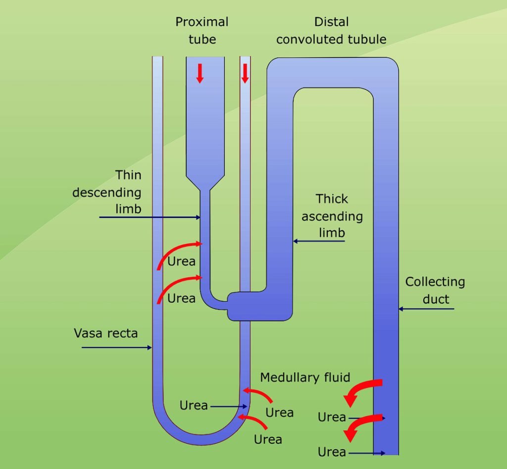
A counter current system of tubules or vessels exists where the inflow of fluid runs parallel to, counter to, and in close proximity to the outflow for some distance. These characteristics are common to the anatomical arrangements of the Loops of Henle and vasa recta. In the kidney, two counter current systems operate-
Counter current multiplier– Loops of Henle
Counter current exchanger– Vasa recta
Counter current multiplier– Loops of Henle
The ascending thick limb of the Loop of Henle is permeable to solutes and so the solutes diffuse into the medullary interstitial fluid with retention of water in the tubule, thereby diluting the tubular fluid. This creates a small osmotic gradient between the tubular and peritubular fluids (interstitial fluid). This osmotic gradient is being multiplied vertically by counter current flow in the descending thin limb (permeable for water and not for solutes).
Water diffuses from the lumen of the descending thin limb into the interstitial fluid. Therefore, the tubular fluid of the descending thin limb increases in osmotic concentration as it descends to the inner most region of the medulla. When thin tubular fluid enters the ascending thin limb (permeable for solute and not for water), sodium chloride diffuses readily outward into the inner medullary interstitial fluid and urea diffuses inward into the tubular fluid.
Continued active secretion by the ascending thick limb, concentration of tubular fluid in the descending thin limb and diffusion from the lumen of the ascending thin limb into the medullary interstitial fluid establishes a vertical osmotic gradient. Therefore, each time sodium chloride makes the circuit around the Loop of Henle, this multiples the concentration of sodium chloride in the medulla and hence, Loop of Henle is called as counter current multiplier.
Countercurrent exchanger- Vasa recta
Countercurrent exchanger is a counter current system in which transport between the outflow and inflow is entirely passive. Vasa recta is permeable to water and solutes through out their length.
In the descending limb of the vasa recta, water is drawn by osmosis from the plasma of vasa recta to the hyperosmotic peritubular fluid (created by counter current multiplier) and the solutes diffuse from the peritubular fluid into the vasa recta.
In the ascending limb, solutes diffuse back into the peritubular fluid and water is drawn by osmosis back into the vasa recta. Net result is that the solutes responsible for medullary gradient are mostly retained in the medulla and the vasa recta carry only slightly more solutes than are brought to them.
Blood flow in the vasa recta is sluggish because an increased rate of medullary blood flow results in decreased time required for diffusion of solute from the ascending limb back to peritubular fluid resulting in gradual loss or wash out of medullary gradient. All the excess salt removed from the interstitial fluid has to be replaced to maintain an osmotic gradient.
Recirculation of urea

Recirculation of urea is a mechanism for concentration of urea in the medulla. Urea is concentrated in collecting tubule and diffuses through the walls of collecting tubule into the medullary interstitial fluid. From there, it is reabsorbed in the Loop of Henle and flows with tubular fluid in the ascending limb through distal tubule into collecting tubule and again out of the collecting tubule into the medullary interstitial fluid. Urea circulates several times before it flows into the urine and it causes urea to accumulate in high concentration in medullary interstitium. This counter current multiplier system helps in concentration of urine and also ensures constant excretion of urea when urine output is low.