TABLE OF CONTENTS
Contraction of digital flexor tendon
Contraction of digital flexor tendon is associated with postural and foot changes, lameness, and debility. They may be congenital and therefore identified in newborn foals or acquired at an older age.
Etiology
Uterine malposition, teratogenic insults (arthrogryposis), and genetic defects have been either implicated or proved to cause contracted limbs in newborn foals.
Chronic pain is the most common cause of acquired tendon contracture. Pain can arise from physitis, osteochondrosis, degenerative joint disease, pedal bone fracture, or soft-tissue wounds and infection. Pain induces reflex muscle contraction with shortening of the flexor musculotendinous units. The horse walks on its toes or knuckles in the fetlocks or occasionally the pastern joint. Nutritional errors referable to problems associated with bone growth (ie, osteochondrosis and physitis) are intimately associated with the syndrome and must be addressed as part of treatment.
Clinical signs
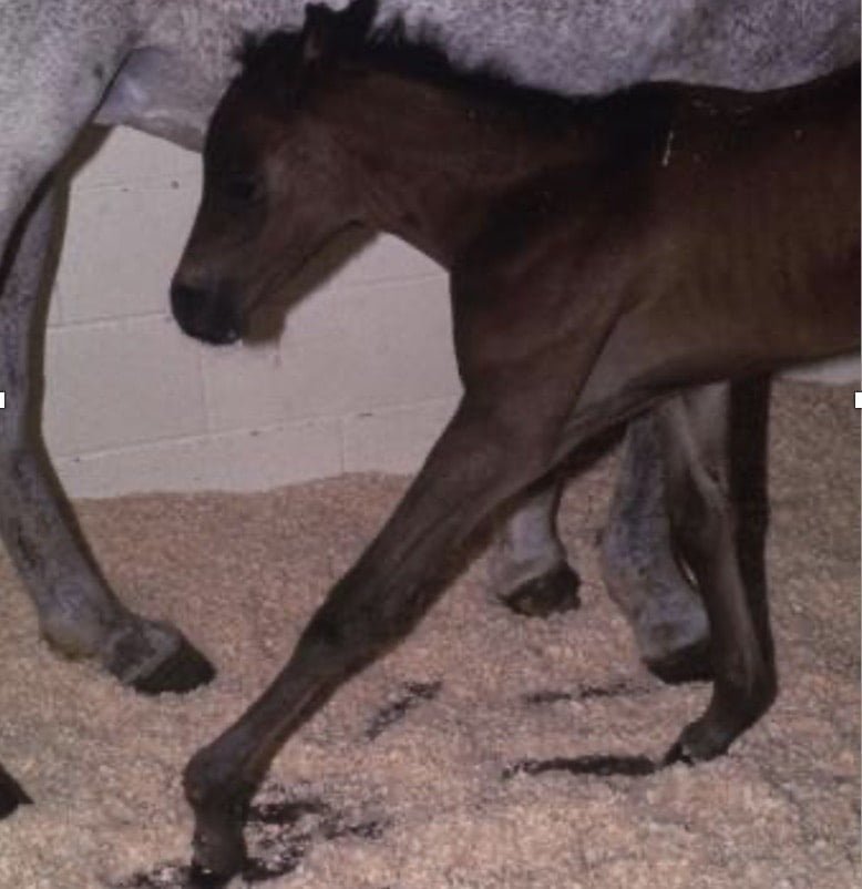
- Signs vary widely in newborn foals. Some cannot stand; some attempt to walk on the dorsum of their fetlocks, and others can stand but knuckle in the fetlocks or carpi.
- One foal may improve spontaneously, yet another, seemingly healthy at birth, may become progressively worse.
- In older foals, onset tends to be rapid; such animals may walk around on their toes with their heels off the ground.
- A slower onset is characterized by an upright hoof with an elongated heel and concave toe. Physitis frequently is evident in these horses.
- Involvement of both forelimbs is common, with a tendency to be worse in one leg.
- Toe abscesses are a frequent complication of the hoof and locomotion changes, and they add to the pain and deformity.
- Older horses (1–2 yr old) commonly knuckle in the metacarpophalangeal joints.
- Yearlings usually are more severely affected and more difficult to treat than younger animals.
- It is important to attempt to identify any underlying bone or joint disease, but this is often difficult and may have resolved.
Treatment
- Mild cases in newborn foals often require no treatment.
- More severe cases require supportive therapy, and it is essential to correct failure of passive transfer of immunity if the foal has not been able to nurse adequately.
- Use of splints necessitates careful fitting and management, because rubbing sores are common and can be severe.
- Casts are generally safer if used only for short periods (5–7 days).
- High-dose oxytetracycline therapy is commonly used (40–60 mg/kg).
- Early acquired cases in older foals and weanlings can be managed conservatively with nutritional correction, proper hoof trimming, and analgesia; however, once the deformity is present for >1 wk, this is rarely successful.
- Surgical treatment can be simple or complex, depending on the degree of involvement.
- Desmotomy of the accessory ligament of the deep digital flexor tendon (inferior check desmotomy) is the most successful and commonly used procedure for flexural deformity of the distal limb and does not interfere with future performance.
- Superior check ligament desmotomy may be included for horses with fetlock deformities.
- For carpal deformities, sectioning of the tendons of insertion onto the ulnaris lateralis and flexor carpi ulnaris is performed.
- In hindlimbs, tenotomy of the medial head of the deep digital flexor is performed, because the inferior check ligament is often vestigial.
- In severe cases, tenotomy of the deep digital flexor tendon can be used as a salvage procedure.
- Nutritional correction, proper foot trimming, and analgesia are integral to recovery when surgery is performed.
- The prognosis is fair to good for horses diagnosed early and managed properly.
Contracted flexor tendons
Contracted flexor tendons are probably the most prevalent abnormality of the musculoskeletal system of newborn foals and calves. An autosomal recessive gene causes this condition. In utero positioning may also affect the degree of disability.
At birth, the pastern and fetlocks of the forelegs and sometimes the carpal joints are flexed to varying degrees due to shortening of the deep and superficial digital flexors and associated muscles. A cleft palate may accompany this condition in some breeds. Slightly affected animals bear weight on the soles of the feet and walk on their toes. More severely affected animals walk on the dorsal surface of the pastern and fetlock joint. If not treated, the dorsal surfaces of these joints become damaged, and suppurative arthritis develops. Rupture of the common digital extensor can occur as a sequela. This condition should be differentiated from arthrogryposis.
Mildly affected animals recover without treatment. In moderate cases, oxytetracycline may be administered to relax the flexor muscles, and a splint can be applied to force the animal to bear weight on its toes. The pressure from the splint must not compromise the circulation, or the foot may undergo ischemic necrosis. Frequent manual extension of the joints, attempting to stretch the ligaments, tendons, and muscles, helps treat these intermediate cases. Severe cases require tenotomy of one or both flexor tendons. A bandage cast may sometimes be indicated. Extreme cases may not respond to any treatment.