TABLE OF CONTENTS
Classical Swine Fever (Hog Cholera)
Swine fever (CF) or Classical Swine Fever (CSF) or Hog Cholera or European swine fever is a highly contagious, often fatal disease of pigs clinically characterized by high body temperature, lethargy, yellowish diarrhea, vomiting and purple skin discolouration of ears, lower abdomen and legs.
Classical Swine Fever (Hog Cholera) is potential to cause devastating epidemics particularly in countries that are free of the disease.
The disease was widespread in South African countries, Europe, China and Japan and prevalent in moderate to severe proportion in India, Myanmar, Nepal, Pakistan, Bangladesh and Philippines.
Etiology
- Classical Swine Fever (Hog Cholera) is caused by genus Pestivirus belongs to family Flaviviridae, it is a small enveloped RNA virus; contain only one serotype, although minor antigenic variability between the strain.
- The virus has antigenic relationship with bovine viral diarrhea virus.
- The virus is moderately fragile and does not survive in an environment.
- However, the virus can survive for prolonged periods in a moist, protein-rich environment such as pork tissue or body fluids particularly if kept cold or frozen, in frozen pork meat the virus can survive for several years.
- The virus is inactivated at pH < 3.0 or > 10.
Epidemiology
- CSF has a worldwide distribution.
- The disease is endemic in central and south America, Asia, Australia, Neweland, Canada, South African countries, Europe, China and Japan, India, Myanmar, Nepal, Pakistan, Bangladesh and Philippines.
- In India this disease was first reported in West Bengal in the year 1961.
- Swine are the natural host of the virus.
- All breeds and all age group of pigs are susceptible to this infection.
- Wild pigs often remain as inapparent carrier of the virus.
- Young pigs are more susceptible than the adult pigs.
Risk factors
- Accidental introduction of CSFV into herds through illegally imported pork meat or pork products.
Source of infection
- Infected live animals can shed the virus in oronasal and ocular secretions, urine, faeces and semen before onset of clinical illness.
- Uncooked pork products.
- In Europe, CSF is endemic in wild boar population; infected wild boars are a source for CSF outbreaks among domestic pigs.
- The probable source for wild boar is contaminated garbage or spill over from the infected pigs
- Untreated swill feeding.
Transmission
- Ingestion of garbage containing uncooked pork products.
- Infection is transmitted readily by direct and indirect contact with an infected animals.
- Inhalation is also a possible portal of entry.
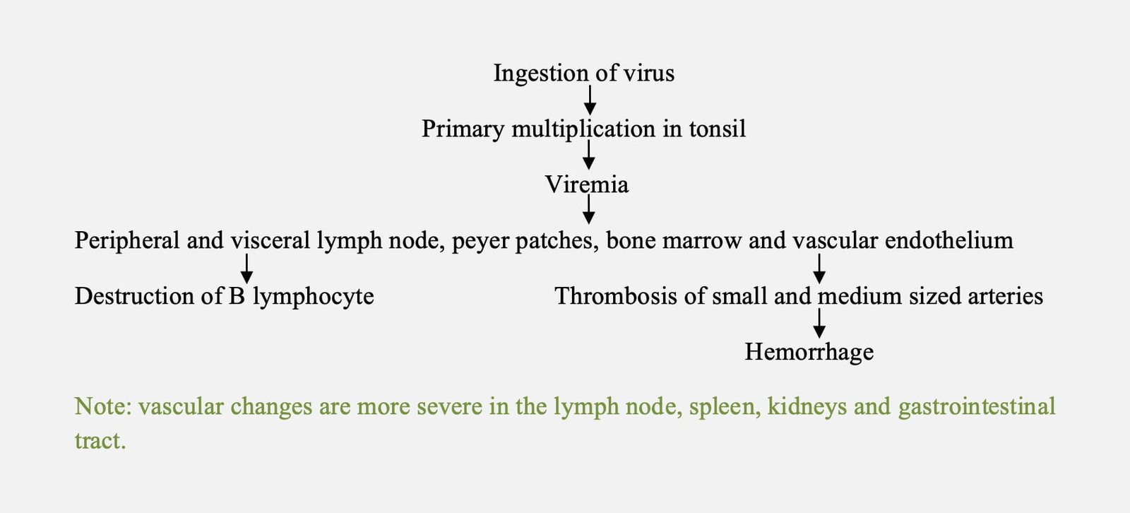
Pathogenesis
Clinical manifestation
- Incubation period is 3-7 days.
- Clinical signs vary with the strain of CSFV, age and breed of the animals.
- The disease may appear in three clinical forms.

Per acute form
- Most commonly noticed in young pigs.
- Disease terminates fatally within about 24 hours of developing disease.
- No appreciable clinical manifestations except high rise of temperature and erythematous patches in the non-hairy parts of the skin.
Acute form
- High fever (105-107o F).
- Huddling, weakness, drowsiness, anorexia and conjunctivitis.
- Disinclination to move, when forced do move, swaying movement of hindquarters often progress to posterior paresis.
- Constipation is followed by intermittent watery diarrhoea.
- Some pigs may vomit yellow bile containing fluid.
- Skin is hyperaemic or may develop haemorrhages on the abdomen, inner thighs, ears or purple cyanotic discolouration on the snout, ears or tail.
- Pigs with acute classical swine fever often die within 1-3 weeks, and convulsions may occur in the terminal stages.
- Convulsions may be accompanied by loud squealing may occur constantly or interval of several hours.
- Tremor of body and limb muscle.
Subacute and chronic form
- The symptoms are similar to acute form, but signs are less severe.
- Poor reproductive performance may be the only sign in some breeding herds infected with less virulent strains.
- Sows in these herds may abort or give birth to stillborn, mummified, malformed, weak or dead piglets.
Necropsy Findings
- Petechial or ecchymotic hemorrhages can often be seen on serosal and mucosal surfaces of the kidney (Turkey egg appearance), urinary bladder, epicardium, epiglottis, larynx, trachea, intestines, subcutaneous tissues and spleen.
- Splenic infarcts (raised, dark, wedge-shaped lesions) are seen occasionally.
- Necrotic foci or “button” ulcers may be found in the intestinal mucosa (colon), epiglottis and larynx.
- Straw colour fluid may be found in the peritoneal and thoracic cavities and the pericardial sac.
- Non suppurative encephalitis.
- In congenitally infected piglets, common lesions include cerebellar hypoplasia, thymic atrophy, ascites, and deformities of the head and legs. Edema and petechial hemorrhages may be seen in the skin and internal organ.
Sample collection
- Live animals – Whole blood and swab from tonsil during fever.
- Dead animal – tonsils, pharyngeal and mesenteric lymph nodes, spleen, kidneys, distal ileum or colon.
Diagnosis
- Based on clinical signs and lesion.
- Marked leukopenia.
- Virus isolation can be carried out in porcine cell lines using homogenates of spleen and tonsil.
- RT-PCR- Most commonly used for detection of viral nucleic acid.
- Loop-mediated isothermal amplification (RT-LAMP) assays have also been published.
- CSFV antigen can be detected by FAT or ELISA.
- Detection of antibodies in serum by virus neutralization test, fluorescent antibody virus neutralization test [FAVN] and neutralizing peroxidase–linked assay [NPLA] tests) and various ELISA test.
Differential diagnosis
- Salmonellosis
- Erysipelas
- Pasteurellosis
- African swine fever
- Anticoagulant poisoning
Treatment
- There is no specific treatment, other than supportive care.
- Hyperimmune serum may be given at dose of 50-150 ml per animal.
- Simultaneous serum- vaccination method.
Prevention and control
Vaccination of pigs with live attenuated vaccine at the age of 1-2 months and booster at 6 months of age and followed by annual revaccination every year.
Endemic area
- Purchase of animals from CSF free herd.
- Quarantine of newly purchased animals for 4 months.
- Testing of animals in quarantine before allowing to the rest of the herd.
Epidemic area
- Eradication by slaughter of all- in contact and infected animal.
- Emergency vaccination can be used for control of the disease in the event of severe outbreak with either Modified live vaccine (of either lapinized strain or cell culture adopted strain) or marker vaccine.
- In wild boar outbreak, emergency vaccination using bait with modified live vaccine.
- Slaughtered pigs are disposed of by burning.
- Restriction of animal movement.
- Disinfection of all vehicle used for transportation, , all pens and premises and utensils must be disinfected with 5% cresylic acid or sodium hydroxide solution.
- Boiling of all contaminated clothing.
- Feeding of boiled garbage.
- Surveillance of animals.
- Avoid contact of domesticated pigs with wild pigs.

