TABLE OF CONTENTS
Cerebellum
Cerebellum is encased in cerebellar hemisphere and is located the back of the brain. Responsible for motor coordination by integrating sensory inputs from receptors located from muscles eyes and ears with motor orders of the fore brain.
It maintains equilibrium and posture.
Present in the anterior part of the brainstem, dorsal to the pons and medulla, and caudal to the cerebral cortex so called as little brain. It contains many neurons.
It is present in all mammals. Jawless vertebrates lack cerebellum.
It is concerned with proper positioning of the body in the space. It maintains subconscious coordination of the movement and coordinates fast phasic motor activity.
Cerebellum is extensive in birds where they need to balance while flying.
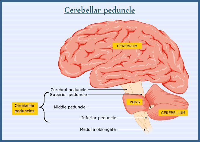
The anatomy of the cerebellum is as follow- Made up of outer grey matter layer known as the CEREBELLAR CORTEX and White matter of cerebellum is represented by stalks known as CEREBELLAR PEDUNCLE.

- Three pairs of the cerebellar peduncle functions as sensory and motor fibers to and from cerebellum and through which it is attached to the brainstem.
- Brachium conjunctivum- to thalamus and mesencephalon. It is the rostral cerebellar peduncle.
- Brachium pontis- to pons. It is the middle cerebellar peduncle.
- Restiform body-to medulla and spinal cord. It is the caudal cerebellar peduncle.

Group of cerebellar nuclei known as subcortical nuclei is embedded in the cerebellar white matter, whose axons leave the cerebellum. They are dentate nucleus, globus emboliformis and fastigial nucleus.
Input to cerebellum
Represented by group of fibers namely Mossy fibers and Climbing fiber axons They use excitatory neurotransmitter to cause excitatory post synaptic potential in the subsequent cells of the cerebellar cortex. The climbing fiber axons exhibit one to one relationship with Purkinje cells.
These fibers carry information from higher motor centres and peripheral sensory receptors.
Both climbing and Mossy sensory fibers are excitatory. When peripheral receptor or CNS is stimulated, it causes activation of these two fibers. The CNS organization facilitates early excitation of Mossy fibers 5-10 millisecond prior to the excitation of climbing fibers. Hence rapid initiation of the cerebellar cortex activity is appreciated.
Excitation of the granule cells and Golgi neurons of cerebellar cortex by Mossy fibers.
Depolarization of dendrites of Purkinje cells, outer stellate, basket cells, and Golgi neurons through parallel fibers from excited granule cells.
Mossy fibers influence the duration of discharge of impulse in the granule.
Inhibition of Golgi neurons are potentiated by stellate and basket cells by way of parallel fibers.
- Degree of inhibition is inversely related to the excitation of climbing fibers that reaches the cerebellar cortex and efferents of the Purkinje cell reflect the cortical activity.
- Cerebellar functions are mediated through disfacilitation of the structures of CNS.
- α-RIGIDITY: It is the rigidity that results from hyper excitation of the α-motor neurons of the spinal cord.
- γ-RIGIDITY: It is the result of the hyper excitation of the γ-motor neurons of the spinal cord.
In birds, cerebellum is concerned with regulation of spinal cord and brain stem mechanism to help in flying and maintenance of equilibrium. Cerebellar pathology is associated with persistent rigidity of the antigravity muscles.