TABLE OF CONTENTS
Canine and Feline Heartworm Disease
Canine and feline heartworm disease is of worldwide significance. But can be prevent by regular deworming and ectoparasiticides that used widely.
Large breed dogs appear to be more susceptible to infection than small breeds, and cats appear to be somewhat resistant to the disease (mosquito bites are less frequent in cats).
Canine Heartworm Disease
The canine heartworm disease is spread by many different species of mosquitoes. Male dogs are more frequently infected compared with female dogs (4:1), and outdoor dogs are more likely to become infected than indoor dogs. The average age at which infections are detected is between 3 and 8 years.
Etiology
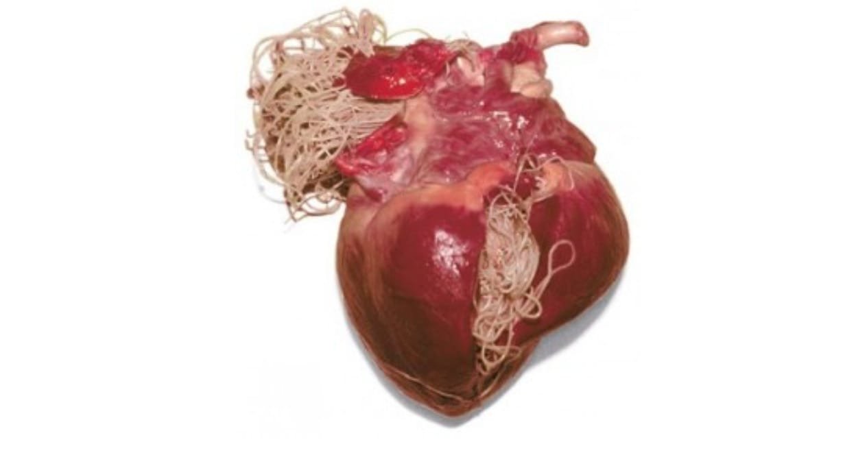
Canine Heartworm Disease caused by a parasitic worm called Dirofilaria immitis.
Pathophysiology & Parasitology
The female mosquito serves as the intermediate host by obtaining a blood meal containing the microfilaria of Dirofilaria immitis from an infected dog.
These microfilaria develop in the mosquito within 2 to 2.5 weeks and are then injected into the skin of another dog through a bite. The infective larvae migrate within the skin of the new host for about 100 days. Young adults (L5 stage) enter the vasculature and migrate to the pulmonary artery, where they mature into adults. Approximately 6 months after the initial bite, microfilaria can be detected in the blood of the host dog.
Disease severity is partially related to the number of adult heartworms. The presence of adult worms in the pulmonary artery damages the endothelial lining of the vessel and increases the permeability, allowing fluid and proteins to leak into the perivascular tissue.
Clinical Signs
- Most dogs are asymptomatic, and infections are discovered on routine screening during yearly examinations
- Cough, dyspnea
- Exercise intolerance
- Haemoptysis (coughing up blood)
- Signs of right sided heart failure
Diagnosis
The physical presence of the parasites results in right-sided heart enlargement (blockage of the right outflow tract) and pulmonary hypertension.
Heartworm disease is easily detected using immuno diagnostic tests that utilize monoclonal antibodies to heartworm uterine antigen. Microfilaria can be detected using filter techniques, the Knott test, or by simply observing a drop of whole blood under a microscope, although these methods may not detect the organisms in as many as 25% of infected dogs.
- Positive antigen test
- Positive concentration test
- Radiography: Evidence of pulmonary changes consistent with heartworm disease: right ventricular enlargement, increased prominence of pulmonary artery, enlarged lobar arteries, increased perivascular pattern
- Echocardiography: adult worms can be seen in the pulmonary artery and sometimes in the right heart
Treatment
Treatment previously involved the removal of the adult worms by the use of thiacetarsamide (Caparsolate), which is no longer available. Melarsamine dihydrochloride (Immiticide, Merial or Diroban, Zoetis) is now the drug of choice for treatment. Animals are prescribed prophylaxis therapy given monthly.
If treatment is elected, the animal should have a pre treatment laboratory workup, which includes a minimum of a complete blood cell count, serum chemistries, and chest radiography.
- Thiacetarsamide: no longer used; serious side effects seen with its use (toxicity occurs in approximately 10% to 15% of cases; signs include bilirubinuria, vomiting, anorexia, lethargy, and icterus)
- Melarsomine dihydrochloride: given at 24-hour intervals. Injections should be made deep into the lumbar muscles
Prevention
- Selamectin
- Ivermectin
- Milbemycin oxime
- Doxycycline
Microfilaria have a symbiotic parasite, Wolbachia, which may be killed by the use of doxycycline. In turn, the death of the parasite adversely affects the adult heartworm. Studies have not been done on the efficacy of this type of therapy.
Feline Heartworm Disease
Cats are also at risk for heartworm infection.
Etiology
Feline heartworm disease is caused by a worm parasite called Dirofilaria immitis.
Cats are somewhat resistant to D. immitis infection, having few adult worms, which are eliminated from the host within 2 years. Outdoor male cats are most at risk for infection. The mean age of diagnosis is between 3 and 6 years.
Clinical Signs
Symptoms in cats differ from those in dogs. Sudden death of asymptomatic cat is fairly frequently seen.
- Coughing
- Dyspnea
- Vomiting
- Anorexia and weight loss
- Lethargy
- Right sided CHF
- Sudden death or acute development of neurological signs
Most symptoms relate to the respiratory system (cough, dyspnea) or the gastrointestinal tract (vomiting, anorexia, lethargy). Acute pulmonary embolism occurs with affected cats demonstrating severe dyspnea, weakness, and anorexia. Ataxia, blindness, and seizures may also be seen.
Diagnosis
It has been difficult to diagnose the disease in cats because they are usually negative for microfilaria, and the canine heartworm antigen tests are inadequate for detecting the disease in cats.
- Feline heartworm antibody immuno diagnostic test (can be done in-house); detection of antibody is missed with this test in many cats
- Feline heartworm antigen immuno diagnostic test: results depend on the sex and number of adult heartworms present
- Radiology: signs are similar to those in the canine but are more difficult to interpret
- Echocardiography: should be done in all cases; will see adult worms in the pulmonary artery
Treatment
Treatment regimens are controversial. In most cases, treatment involves supportive care while the cat eliminates the parasite.
- Thiacetarsamide and immiticide both have toxic consequences in the cat and may be fatal.
- Microfilariacide: not needed in cats because of lack of microfilaria
- Cage rest
- Cortisone may be used to decrease the inflammatory component of the disease
Prevention
Prevention is advised and is now available for cats at risk. Milbemycin oxime or ivermectin given monthly is very effective.

