TABLE OF CONTENTS
Anatomy of the Bony Pelvis in Animals: Sacrum, Coccygeal Vertebrae, and Pelvic Inlet/Outlet Diameters
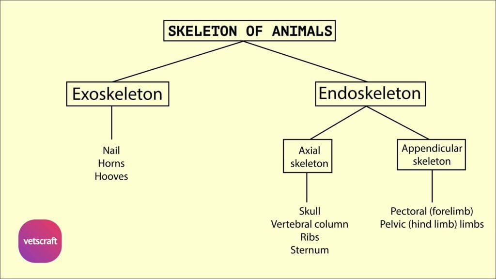
The bony pelvis of domestic animals consists of the sacrum, the first three coccygeal vertebrae, and two os coxae formed by the ilium, ischium, and pubis.
The sacrum shows species-specific variations in the number of segments, shape, surface features, and degree of vertebral fusion among animals such as the mare, cow, buffalo, sheep, sow, and dog.
Coccygeal vertebrae also vary in number, structure, and the presence of features like spinous processes and arterial grooves.
The bony pelvis, or bones of the hip, consist of the sacrum, the first three coccygeal vertebrae, and two os coxae. Each os coxae is formed by the ilium, ischium, and pubis.
Sacrum
The sacrum is a triangular bone formed by the fusion of sacral vertebrae, connecting the spine to the pelvis. It articulates with the ilium on either side to form the sacroiliac joints, contributing to the stability of the pelvic girdle.
Comparison of Sacrum Between Mare and Cow
| S. No. | Mare | Cow |
|---|---|---|
| 1 | It consists of 5 sacral segments, a base, and an apex. | It consists of 5 sacral segments. |
| 2 | It is shorter than in the cow. | It is longer than in the mare. |
| 3 | It is triangular in shape. | It is roughly triangular in shape. |
| 4 | The dorsal surface presents 5 sacral spines with tuberous summits and 4 pairs of dorsal sacral foramina. | The dorsal surface presents fused spines forming the median sacral crest. The articular processes fuse to form the lateral sacral crest and 4 pairs of dorsal sacral foramina. |
| 5 | The ventral surface bears 4 transverse lines. | The ventral surface presents 4 transverse grooves and 4 pairs of foramina. |
| 6 | The pelvic surface presents transverse lines. | The pelvic surface is concave in both directions and presents a groove for the middle sacral artery. |
| 7 | Smooth notches are present on either side of the body of the 1st sacral segment. | No notches are present on either side of the body of the 1st sacral segment. |
| 8 | The articular surface is directed dorso-laterally, slightly concave along its length. It is rough, irregular, and articulates with the ilium. The portion lateral to the foramina is formed by the fusion of the transverse processes. | The articular surfaces are concave and semicylindrical medially; the processes are large and widely separated. |
| 9 | The alae form the lateral part of the base (wing). The ventral margin of the base projects to form the sacral promontory. | — |
Comparison of Sacrum Between Buffalo and Sheep
| S. No. | Buffalo | Sheep |
|---|---|---|
| 1 | It consists of 5 sacral segments. | It consists of 4 sacral segments. |
| 2 | No vascular groove is present on the ventral aspect. | No vascular groove is present on the ventral aspect. |
| 3 | Spines are fused. | Spines are not fused except for the 1st and 2nd, which may be partially united. |
| 4 | The sacral promontory is flattened dorso-ventrally more than in the cow. | The sacral promontory is not significantly flattened dorso-ventrally. |
| 5 | The 4th and 5th sacral vertebrae are not fused but are laterally attached. | The last segment may remain separate or undergo partial fusion. |
| 6 | The transverse processes of the last segment are not distinct. | The transverse processes of the last segment are distinct and prominent. |
Comparison of Sacrum Between Sow and Bitch (Dog)
| S. No. | Sow | Bitch |
|---|---|---|
| 1 | It consists of 4 sacral segments. | It consists of 3 sacral segments. |
| 2 | The pelvic surface is less curved compared to that of the cow. | It is short, wide, and quadrangular in shape. |
| 3 | Fusion of vertebrae is less complete. | Fusion of vertebrae is complete. |
| 4 | Spines are less developed and commonly absent. | Spines are fused to form a median crest, which is notched. On either side, two tubercles represent the vestiges of the fused articular processes. |
| 5 | The middle of the dorsal surface is flattened, smooth, and presents an opening. | The pelvic surface is deeply concave and has two pairs of foramina. |
| 6 | The anterior processes are very large. | The anterior processes are large with extensive, slightly concave facets. The wings are prismatic and bear a laterally positioned auricular surface on the lower part. |
| 7 | The transverse lines are distinct. | The sacral canal is compressed dorso-ventrally. |
| 8 | — | The transverse processes of the last vertebra project backward. |
Coccygeal Vertebrae
Coccygeal vertebrae, also known as caudal or tail vertebrae, form the skeletal structure of the tail in animals. Their number and morphology vary significantly among species, with differences in length, fusion, presence of spinous processes, and arterial grooves.
Comparison of Coccygeal Vertebrae Between Mare and Cow
| S. No. | Mare | Cow |
|---|---|---|
| 1 | The coccygeal vertebrae (Cy) are 15 to 21 in number, typically 18. | The coccygeal vertebrae (Cy) are 18 to 20 in number. |
| 2 | The spinous processes are short and have a double summit. | — |
| 3 | The 1st vertebra presents a groove on the postero-medial aspect of the body. | It is longer and better developed than in the horse. |
| 4 | The ventral surface has a groove for the coccygeal artery. | Ventrally, it has a median sacral groove for the median coccygeal artery. |
| 5 | The vertebrae are ill-developed. | The first 5 to 6 vertebrae have complete arches and spinous processes. |
| 6 | The bodies of the first 3 vertebrae are flattened dorso-ventrally and constricted in the middle. | In the anterior part of the series, the anterior articular processes do not articulate, and the ventral spines form a groove for the middle coccygeal artery. |
Comparison of Coccygeal Vertebrae Between Sheep and Sow
| S. No. | Sheep | Sow |
|---|---|---|
| 1 | The coccygeal vertebrae (Cy) are 16 to 18 in number, with a possible range from 3 to 24. | The coccygeal vertebrae (Cy) are 20 to 23 in number. |
| 2 | The transverse processes are long and thin. | The first 3 or 4 vertebrae are notably characterized by the presence of functional articular processes. |
| 3 | No haemal processes are present on the ventral surface of the body. | — |
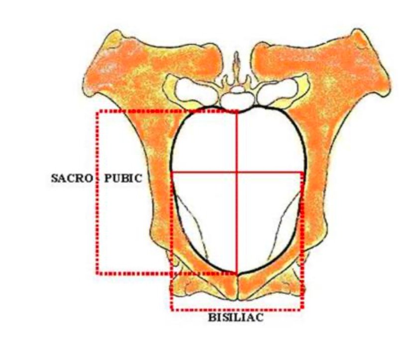
Diameters of Pelvic Inlet
- Sacro-pubic (Conjugate) diameter: It is measured from the sacral promontory to the anterior margin of the pubic symphysis.
- Transverse (Bis-iliac) diameter: Measured at its greatest width just above the psoas tubercle.
- Superior bis-iliac diameter: It is measured at the upper third of the pelvic inlet and receives the greatest width of the fetus at the shoulders in anterior presentation and at the hips in posterior presentation.
- Inferior bis-iliac diameter: It is measured at the lower fourth of the pelvic inlet, at the elbow of the fetus in anterior presentation and at the stifle in posterior presentation.
- Vertical diameter of inlet: It is measured between the anterior end of the pubic symphysis and the articulation of the third and fourth sacral vertebrae.
- Oblique / sacro-iliac / ilio-sacral diameter of inlet: It is measured from the sacro-iliac joint on one side, through the center of the pelvic cavity, to the psoas tubercle on the opposite side. It is intermediate between the sacro-pubic and superior bis-iliac diameters.
Diameter of Pelvic Outlet
- Superio-inferior (Vertical) diameter: It is measured between the summit of the ischial arch and the articular surface of the first and second coccygeal vertebrae.
- Transverse diameter: It is measured between the two ischiatic spines (Chauveau).