TABLE OF CONTENTS
Bicarotid trunk
- The bicarotid trunk continues the brachiocephalic artery after it detaches the right brachial artery.
- It is directed forwards beneath the trachea above the jugular confluence.
- It is related laterally to the vagus and recurrent laryngeal nerves.
- At the thoracic inlet, it bifurcates into two common carotid arteries-right and left.
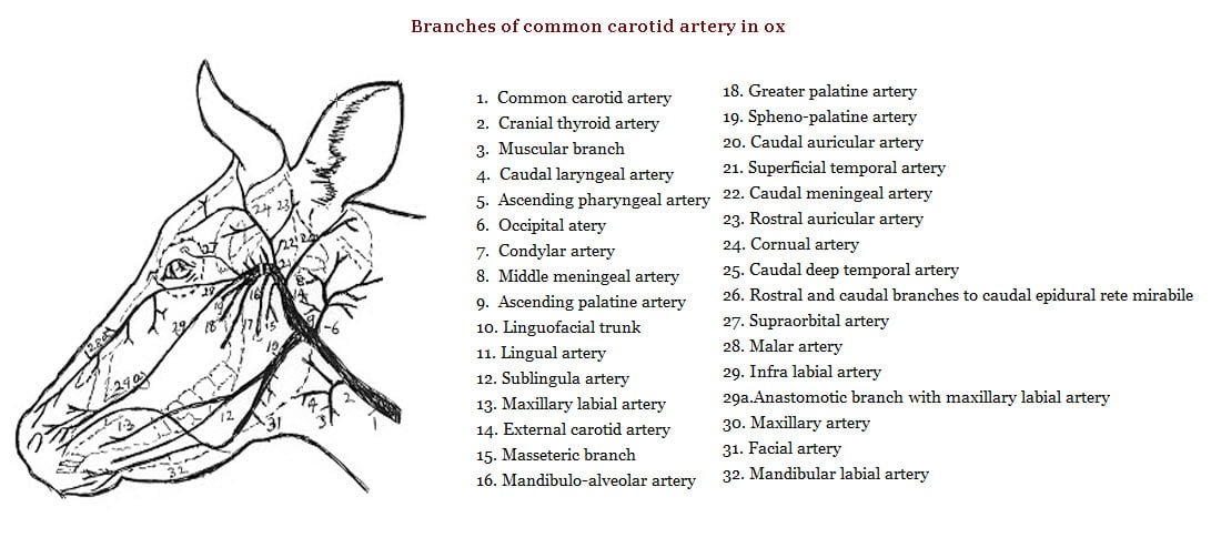
Common carotid artery
- Each common carotid artery passes up obliquely on the side of the neck and diverges from its fellow as it ascends.
- It crosses the trachea obliquely being at first below then lateral to it and finally on its dorsal face at the termination.
- It is related superficially to the internal jugular vein and the tracheal lymph duct, dorsally to the vagosympathetic trunk and ventrally to the right recurrent laryngeal nerve.
- It is separated from the external jugular vein by the sternomastoideus and omohyoideus.
- It is related in its anterior two thirds to the longus colli, rectus capitis ventralis major and omohyoideus dorsally and to the scalenus laterally at the lower third.
- Each artery is deeply placed at its termination and is related to the parotid and mandibular salivary glands laterally and atlantal lymph glands medially.
- The left common carotid artery has the oesophagus deeply ventral to it in the jugular furrow and the left recurrent laryngeal nerve on this side passes ventral to the oesophagus and between it and the trachea and at its termination it is related to the right common carotid artery.
- The common carotid artery terminates at the level of the digastricus by dividing into occipital, external maxillary and carotid arteries.

Collateral branches
- Branches to the trachea and oesophagus.
- The thryo-laryngeal artery is the largest of the collateral branches.
- It arises from the common carotid artery a little in front of the preceding 2″ to 3″ behind its final division.
- It divides into thyroid and laryngeal branches.
- The thyroid branch enters the anterior extremity of the lateral lobe of the thyroid gland and is distributed to it.
- The laryngeal branch divides into pharyngeal and laryngeal branches of which the former is distributed to the muscles of the pharynx and the latter to the muscles of the larynx.
- The posterior thyroid artery when present enters the posterior extremity of the lateral lobe of the thyroid gland.
- The inferior parotid artery to the parotid salivary gland.
- The posterior meningeal artery, which enters the cranium through the foramen lacerum and contributes to the formation of the rete mirabile cerebri.
- Muscular branches to the ventral muscles of neck.

