TABLE OF CONTENTS
Avian Leukosis
Avian Leukosis is also known as Avian sarcoma, Big liver disease, visceral lymphoid leukosis and Lymphomatosis.
The Avian Leukosis sarcoma viruses cause erythroid, lymphoid and myeloid leukosis, a variety of other tumours, such as fibrosarcomas, hemangiomas, nephroblastoma and bone disorder osetopetrosis.
- Lymphoid leukosis- common in layers.
- Myeloid leucosis and erythroid leukosis- common in meat type birds (Broilers).
Lymphoid leukosis is a common neoplasm caused by ALV subgroup A and B. ALV-J has weak ability to cause lymphoid leukosis in layers.
Etiology
- Avian Leukosis is caused by an Alpharetrovius of family Retroviridae.
- The viral structural protein p27 is major group specific antigen encoded by gag gene. Which is common to all viruses of leukosis or Sarcoma group.
- ALSV in chickens are classified into six subgroups (A, B, C, D, E & F) on the basis of differences in their viral envelope glycoprotein gp 85.
- Most of the viruses causing avian leukosis are exogenous. Endogenous ALGVs, which mostly belong to subgroup E and occur commonly in chickens and other avian species are of little or no pathogenicity.
Host affected
- Chicken are natural host for the virus, the virus also isolated from pheasants, partridge and quail.
- Adult bird greater than 16 weeks of week old are commonly affected.
Transmission
- Vertical transmission- most common (from hen to progeny through the egg)
- Horizontal transmission through faeces of infected chicks to susceptible host
- Congenitally infected chicks develop immunological tolerance to the virus and fail to develop neutralizing antibodies and permanently shed the shed organism.
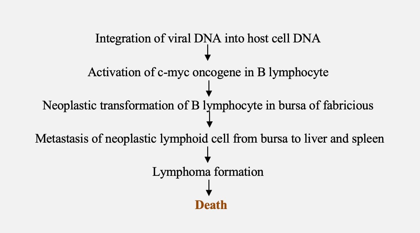
Pathogenesis
- ALV infection in the absence of neoplastic disease can adversely affect egg production traits in layers, including age at first egg production, number, size, fertility and hatchability.
- Broilers: suppress the growth rate
Clinical manifestation
- The incubation period is variable and may be as long as months or years
- Unthriftiness, inappetence, weakness, diarrhoea, dehydration and emaciation
- Pale comb
- Enlarged abdomen is due to tumour growth in liver
- Infected chicken is depressed before death
- Reduced egg production occur in subclinical infection
Necropsy finding
- Diffuse enlargement of liver and nodular tumour is common in liver, spleen and bursa and occasionally found in kidney, gonads and messentry.
- Histopathological examination- uniform infiltration large lymphoblast cells in liver, spleen and bursa.
Sample collection
Buffy coat, egg white, paired sera sample, liver, spleen and bursa of fabricious.
Diagnosis
- Isolation of virus in cell culture
- Histopathological examination
- Cytological examination of May-Grunwald-Giemsa stained impression smear of fresh tumour
- Detection of antigen by RT-PCR
- Detection of antibodies by ELISA
Control
- Eradication of avian leukosis virus from primary breeding flocks
- Breeder flock is evaluated by shedding of virus in albumin of egg by ELISA
- Eggs from shedder are discarded
- Selection of genetically resistance line for breeding
- There is no commercial vaccine
Erythroid leukosis (Erythroblastosis)
- Erythroid leukosis (Erythroblastosis) is caused by ALV- J subgroup
- Meat type bird (broilers) are commonly affected
- Adult chickens are mostly affected
- Anemia with immature RBCs in the blood
- Disease originate from bone marrow
- In Malignant form- the cells remain within the blood vessels throughout the course of the disease
- Erythrostasis in sinusoids of liver, spleen and bone marrow imparting cherry red colour
- Liver and spleen is moderately enlarged
Myeloid leukosis (Myeloblastosis and Myelocytomatosis)
- Myeloid leukosis (Myeloblastosis and Myelocytomatosis) is caused by ALV- J subgroup
- It involves extravascular and intravascular proliferation of myeloid (granulocytic series
- Myeloblastosis – liver and spleen are diffusely enlarged, the liver frequently as a granular, morocco leather appearance
- Myelocytomatosis- tumour is nodular or diffuse have a creamy white colour. The tumour growth occur in liver, spleen, kidney, flat bones of rib, skull, sternum and pelvis
Osteopetrosis
- Osteopetrosis is a sporadic disease that occurs primarily in males
- The shafts of the long bones are thickened, often resulting in a stilted gait
- Affected birds may be anemic and also have lesions as that of lymphoid leukosis.

