TABLE OF CONTENTS
Aural Hematoma
Aural Hematoma is the accumulation of blood between the ear cartilage and the skin either in the internal or external aspect due to trauma. It is common surgical affection of ears in equines, dogs, rabbit and goats.
In breeds of dog with long and pendulous ears. If not treated, It may organises into large scar causing deformity in shape of ear pinna.
Etiology of aural hematoma
Etiology of aural hematoma are inflammations, parasites, allergy, and foreign bodies common physical injury of the ear flap, self inflicted by scratching and head shaking.
Pathophysiology of aural hematoma
Trauma of ear pinna cause venous bleeding at the tip. Bleeding from auricular artery can also cause aural hematoma. Bleeding continuous until the initial pressure equalise the pressure of the bleeding vessels. Additional trauma on haematoma causes further seperation of the tissue and allows haemorrhage to resume. Fibrin is deposited on walls of the haematoma, leaving a sanguinous seroma. With chronicity, fibrosis and contraction thicken and deform the ear. Fibrous reorganisation of the ear and secondary cauliflower contracture can result.
Clinical signs of aural hematoma
Swelling of the flap is more evident on the concave surface.
Treatment of aural hematoma
Needle Aspiration
Needle Aspiration is employed soon after haematoma formation to evacuate the contents and restore tissue apposition. Chance of recurrence and complications are more in this approach.
Placing self retaining drainage tubes
Aural Hematoma in which minimum fibrin is present can be treated by placing a self retaining disposable teat canula through a small stab incision made at the dependant portion of AH near the ear tip. If continuous drainage is allowed for 2-3 wks, resolution is likely.
Drain tube method
Under General Anaesthesia, on lateral recumbency, Plug the ear canal. Make a trianguIar shaped incision at the tip of the bulging under aseptic precautions and drain the contents. Similar type incision can be made at the base of the bulging. Try to remove fibrous adhesions through the incisions using an artery forceps. A flexible fenestrated tube can be passed thr’ the incisions and fixed to the pinnae by sutures for drainage.
Post operative care: Bandage the ear pinna folded around a conical roll of sterile guaze kept on the internal aspect. Remove the bandage after 24 hrs and clean the tube using NS and infuse the cavity with Povidone iodine. Advise the owner to press the pinnae evenly for 2-3 times daily to evacuate the contents as much as possible and to dress the wounds with antiseptics. The ear may be protected by applying an E-Collar for 2-3 wks. The tube can be removed after cutting the sutures at the end of 3rd wk. Systemic antibiotics can be administered for a wk period.
Surgical operation for aural hematoma
Large ,severe or chronic AH are treated by incision to remove fibrinous attachment and suturing the integuments to re-appose the tissue layers.
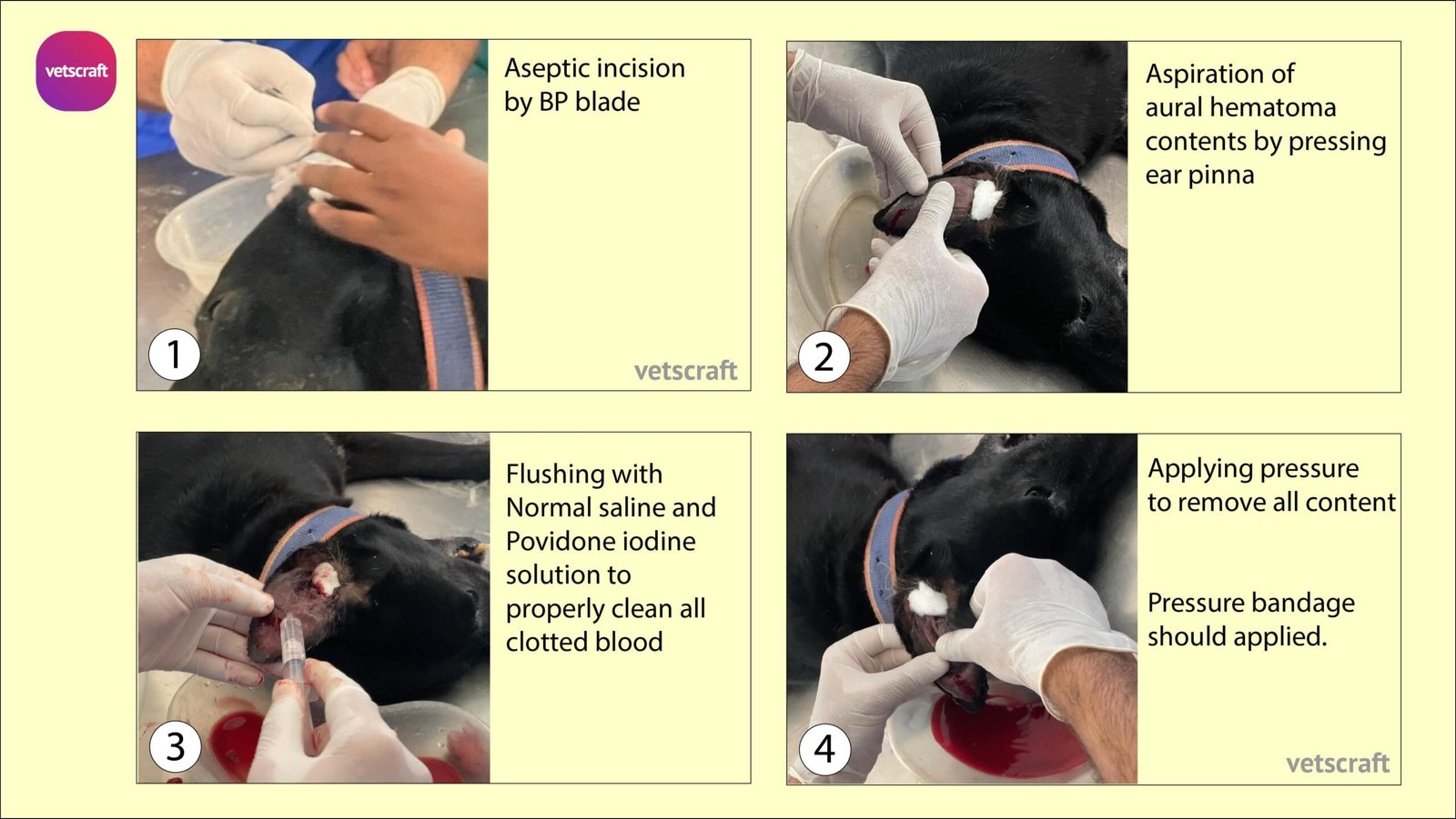
Under General Anaesthesia, Prepare the internal and external aspect of the AH for aseptic surgery. Plug the ear canal. A straight/ cruciate/ S shaped incision is made on inner aspect of pinnae aseptically. Drain the contents, scrape the fibrinous adhesions and smear the lining of the cavity with povidone Iodine.
Button suture can be applied to overcome recurrence of aural hematoma.
Pressure bandage can be applied and animals should be review after 2-3 days for repeat the same procedure. Antibiotics should be advised.