TABLE OF CONTENTS
Anatomy of Atlas Vertebra in Domestic Animals
The atlas is the first cervical vertebra (C1) in animals, situated immediately caudal to the skull. It articulates with the occipital condyles of the skull to form the atlanto-occipital joint, allowing dorsoventral flexion of the head.
Structurally, the atlas is atypical—it lacks a vertebral body and spinous process and instead forms a ring composed of dorsal and ventral arches with prominent lateral wings.
Atlas of Ox
The atlas of the ox is atypical in form and structure. The body and spinous process are absent. It has the form of a ring from which two curved plates, the wings, project laterally.

Ring
The ring encloses a large vertebral foramen and consists of two lateral masses connected by dorsal and ventral arches.
The lateral masses present two deep anterior articular cavities, which receive the occipital condyles; the cavities are partially divided into dorsal and ventral parts by a non-articular area and are separated by a narrow interval below.
The posterior articular surfaces are confluent on the ventral arch, flattened behind, and are continued into the vertebral canal forming an extensive area for the dens of the axis.
The dorsal arch presents a median dorsal tuberosity and is concave ventrally. It is perforated on either side near its anterior margin by the intervertebral foramen, which is connected by a short groove—the alar groove—with another foramen, the alar foramen, which perforates the wing. The anterior border of the dorsal arch is notched. The posterior is thin and shows a central and two lateral notches.
The ventral arch is thicker and less curved than the dorsal. On its lower surface in the median plane is an obtuse eminence, the ventral tubercle, for the tendon of longus colli. The dorsal face has posteriorly a transversely flat articular surface, the fovea dentis, for the dens of the axis. In front of this is a transverse rough area for the ligamentum dentis (odontoid ligament).
Wings
The wings represent the transverse processes of the other vertebrae. Each is a horizontal plate of bone springing from the lateral aspect of the lateral masses and terminating outward in a rough, thick edge. This edge furnishes attachment to the muscles of the head and neck.
The dorsal surface is rough. Between the ventral aspect of the wing and the lateral mass is a cavity—the fossa atlantis. At the bottom of the fossa are two foramina: the anterior, called the alar foramen, perforates the wing and allows passage of the ventral primary branch of the first spinal nerve and branches of vertebral vessels.
The posterior foramen pierces the lateral mass and communicates with the spinal canal. The alar foramen is connected on the dorsal aspect by a short alar groove with the intervertebral foramen.
Comparative Anatomy of Atlas
The atlas vertebra exhibits notable anatomical variations among different animal species, reflecting differences in head movement and structural adaptation.
Sheep and Goat
- The atlas differs chiefly in that the prominence on the dorsal arch is much less developed in the case of sheep and goats.
Horse
- Atlas of horse’s wing slopes downwards and outwards.
- Besides the alar and intervertebral foramina, there is a foramen transversarium at the posterior part of the wing.
- The posterior edge of the dorsal arch does not present the notches as in the ox.
- The posterior articular surfaces are saddle-shaped.

Pig
- The atlas of the pig has a large tuberosity.
- The wing is flattened and bears a posterior tuberosity.
- The foramen transversarium is small or absent.
Dog
- The ventral arch is narrower from before backward and bears a small tubercle posteriorly.
- The dorsal surface of the dorsal arch is strongly convex and rough centrally.
- The wings are horizontal.
- An alar notch is present on the anterior border instead of an alar foramen.
- The foramen transversarium is present.
Rabbit
- Atlas of rabbit is comparatively shorter than that of the ox.
Fowl
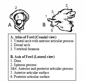
- Atlas of fowl is a ring-like bone.
- The ventral arch presents anteriorly a concave articular area for the single occipital condyle.
- Posteriorly, there are three facets for the axis – one median on the ventral arch and two on the posterolateral aspects of the dorsal arch.