TABLE OF CONTENTS
Angular limb deformities
Angular limb deformities are congenital or acquired skeletal defects, the distal portion of a limb deviates laterally or medially early in neonatal life. In utero malposition, hypothyroidism, trauma, poor conformation, excessive joint laxity, and defective endochondral ossification of the carpal or tarsal and long bones have been implicated. One to four limbs may be affected, depending on the severity of the condition.
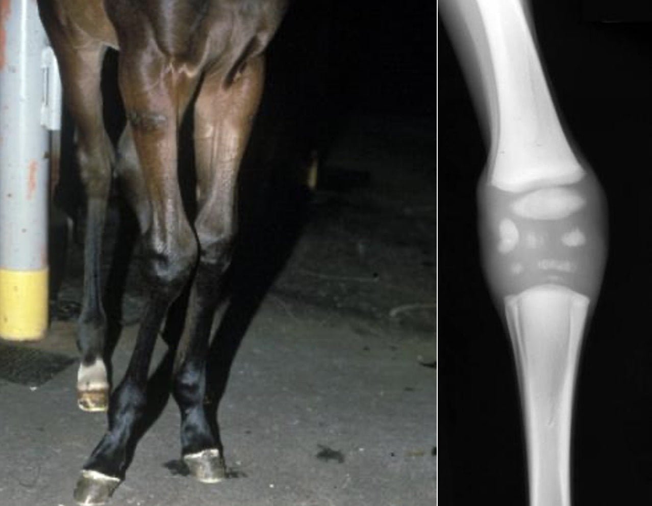
The carpus is affected most frequently, but the tarsus and fetlocks are occasionally involved. The deviation is obvious but varies in severity. A lateral deviation (valgus) of up to 6° of the distal portion of a limb may be regarded as normal. Most foals are asymptomatic, but lameness and soft-tissue swelling can accompany severe deviations. Outward rotation of the fetlocks invariably accompanies carpal valgus. Foals with defective ossification of the carpal cuboidal bones or excessive joint laxity are frequently lame as the legs become progressively deviated. Affected limbs must be palpated carefully to detect ligament laxity and specific areas that may be painful.
Diagnosis
Diagnosis should include a precise determination of the site and cause for the deviation. The distal radial metaphysis, physis, epiphysis, or cuboidal bones may be the site of deviation. Radiography helps detect physeal flaring, epiphyseal wedging, and deformation of carpal bones. Mildly affected foals frequently improve spontaneously.
Treatment
Treatment depends on the severity of the condition and tissues affected. Excessive joint laxity, with or without cuboidal carpal bone involvement, requires tube casts or splints. The fetlock and phalangeal region should not be included in the casts, which should protect the weak joint from trauma but allow restricted exercise to maintain tendon and ligament tone. Such limb support may be required for as long as 6 week.
Physeal and epiphyseal growth disturbances are also amenable to surgical correction through hemicircumferential transection and periosteal elevation of the distal radius on the concave side of the defect or through transphyseal bridging of the physis on the convex side. These surgeries must be performed before the physeal growth plates close (as early as 2–4 mo of age for fetlocks), and success depends on continued growth and development of the bones. Sequential examinations and radiographs are necessary to follow spontaneous improvement or to establish a need for surgery.
Prognosis
Without treatment, the prognosis for severe carpal valgus is poor. The conformational anomaly leads to early degenerative joint disease. Likewise, deformity of the cuboidal carpal bones contributes to a poor prognosis. However, with early detection, careful evaluation, and proper surgical treatment, most foals respond favorably.